Application
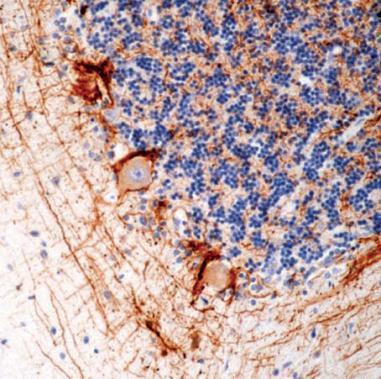
IHC Image using - Detection of 200kDa + 70kDa Neurofilament by IHC in Human Cerebellum| Product Name | NF-H + NF-L antibody |
|---|---|
| Antibody Type | Primary Antibodies |
| Product description | Mouse Monoclonal antibody [2F11] to NF-H + NF-LNeurofilaments can be defined as the intermediate or 10nm filaments found in specifically in neuronal cells. In the electron microscope neurofilaments appear as 10nm diameter fibres of indeterminate length which generally have fine wispy protrusions from their sides. They are found particularly abundantly in axons of large projection neurons. They probably function to provide structural support for neurons and their synapses and to support the large axon diameters required for rapid conduction of impulses down axons. They are composed a mixture of subunits which usually include the three neurofilament triplet proteins known as NFL, NFM and NFH. Neurofilaments may also include smaller amounts of peripherin, alpha internexin, nestin and in some cases vimentin. Antibodies to the various neurofilament subunits are very useful cell type markers since the proteins are among the most abundant of the nervous system, are expressed only in neurons and are biochemically very stable. |
| Immunogen | Purified neurofilaments from human brain |
| Clonality | Monoclonal |
|---|---|
| Isotype | IgG1 |
| Clone Number | 2F11 |
| Host Species | Mouse |
| Tested Applications | IHCIHC-PIPWB |
| For IHC-P: Use at a dilution of 1:25-1:50 in an ABC method. Incubate for 30 minutes at RT. For IP: Use at a dilution of 1:50. for WB: Use at a dilution of 1:50-1:100. Optimal dilutions/concentrations should be determined by the end user.: |
|
| Species Reactivity | HumanMouseDogPigRatCatHorseOpossumRabbitBovine |
| Concentration | 1mg/ml |
| Purification | Affinity purified |
| Gene Synonyms | Neurofilament light polypeptide 70kDa Neurofilament triplet L protein NEFL NFL 70kDa neurofilament protein 200kDa + 70kDa Neurofilament Neurofilament triplet H protein CMT1F NFH Neurofilament light polypeptide70kDa NF70 KIAA0845 NEFH |
|---|---|
| Target | NF-H + NF-L |
| Tissue Specificity | Detects the 70 and 200 kD component of the polypeptide subunit. It stains neurons, neuronal processes in central and peripheral nerves. reacts with some neuroblastomas and gangliomas but not with other non-neuronal tumors. |
| Cellular Localization | Cytoplasmic |
| Light Chain Type | kappa |
Application
IHC Image using - Detection of 200kDa + 70kDa Neurofilament by IHC in Human Cerebellum| Positive Control | Brain |
|---|---|
| Application Notes | For IHC-P: Use at a dilution of 1:25-1:50 in an ABC method. Incubate for 30 minutes at RT. For IP: Use at a dilution of 1:50. for WB: Use at a dilution of 1:50-1:100. Optimal dilutions/concentrations should be determined by the end user.: |
| Form | Liquid |
|---|---|
| Storage Instructions | Keep as concentrated solution, aliquot and store at 4°C. Do not freeze. |
| Storage Buffer | Tissue culture supernatant containing sodium azide |


