Application
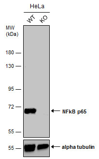
Wild-type (WT) and NFkB p65 knockout (KO) HeLa cell extracts (30 µg) were separated by 7.5% SDS-PAGE, and the membrane was blotted with NFkB p65 antibody diluted at 1:500. The HRP-conjugated anti-rabbit IgG antibody was used to detect the primary antibody.Application
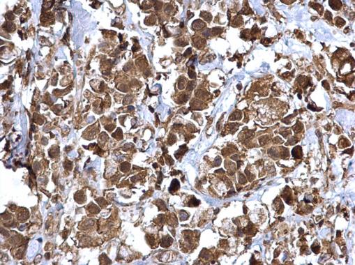
NFkB p65 antibody detects NFkB p65 protein at cytosol and nucleus on human breast carcinoma by immunohistochemical analysis. Sample: Paraffin-embedded human breast carcinoma. NFkB p65 antibody dilution: 1:500.Application
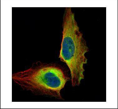
Confocal immunofluorescence analysis (Olympus FV10i) of paraformaldehyde-fixed U2OS, using NFkB p65 antibody (Green) at 1:500 dilution. Alpha-tubulin filaments were labeled with (Red) at 1:2000.Application
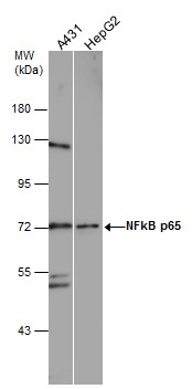
Various whole cell extracts (30 µg) were separated by 7.5% SDS-PAGE, and the membrane was blotted with NFkB p65 antibody diluted at 1:1000. The HRP-conjugated anti-rabbit IgG antibody was used to detect the primary antibody.Application
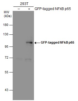
Non-transfected (–) and transfected (+) 293T whole cell extracts (30 µg) were separated by 7.5% SDS-PAGE, and the membrane was blotted with NFkB p65 antibody diluted at 1:5000. The HRP-conjugated anti-rabbit IgG antibody was used to detect the primary antibody.Application
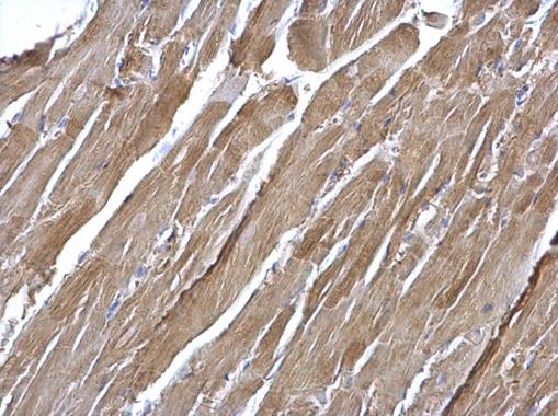
NFkB p65 antibody detects NFkB p65 protein at cytosol on mouse heart by immunohistochemical analysis. Sample: Paraffin-embedded mouse heart. NFkB p65 antibody dilution: 1:500.| Product Name | NFkB p65 antibody |
|---|---|
| Antibody Type | Primary Antibodies |
| Product description | Rabbit Polyclonal antibody to NFkB p65 (v-rel reticuloendotheliosis viral oncogene homolog A (avian))NFKB1 (MIM 164011) or NFKB2 (MIM 164012) is bound to REL (MIM 164910), RELA, or RELB (MIM 604758) to form the NFKB complex. The p50 (NFKB1)/p65 (RELA) heterodimer is the most abundant form of NFKB. The NFKB complex is inhibited by I-kappa-B proteins (NFKBIA, MIM 164008 or NFKBIB, MIM 604495), which inactivate NFKB by trapping it in the cytoplasm. Phosphorylation of serine residues on the I-kappa-B proteins by kinases (IKBKA, MIM 600664, or IKBKB, MIM 603258) marks them for destruction via the ubiquitination pathway, thereby allowing activation of the NFKB complex. Activated NFKB complex translocates into the nucleus and binds DNA at kappa-B-binding motifs such as 5-prime GGGRNNYYCC 3-prime or 5-prime HGGARNYYCC 3-prime (where H is A, C, or T; R is an A or G purine; and Y is a C or T pyrimidine).[supplied by OMIM] |
| Immunogen | Recombinant protein encompassing a sequence within the center region of human NFkB p65. The exact sequence is proprietary. |
| Clonality | Polyclonal |
|---|---|
| Isotype | IgG |
| Host Species | Rabbit |
| Tested Applications | ICC/IFIHC-PWBChIP |
| *Optimal dilutions/concentrations should be determined by the researcher.
: |
|
| Species Reactivity | HumanMouse |
| Predicted Reactivity | Rat, Pig, Chimpanzee, BovineRat (99%), Pig (98%), Chimpanzee (99%), Bovine (98%) |
| Concentration | 1mg/ml |
| Purification | Affinity purified |
| Gene Synonyms | MGC131774 antibody NFKB3 antibody p65 antibody RELA antibody nuclear factor NF-kappa-B p65 subunit antibody transcription factor p65 antibody NF-kappa-B p65delta3 antibody nuclear factor of kappa light polypeptide gene enhancer in B-cells 3 antibody |
|---|---|
| Molecular Weight(MW) | 60 kDa (note) (4) |
| Cellular Localization | Nucleus , Cytoplasm |
Application
Wild-type (WT) and NFkB p65 knockout (KO) HeLa cell extracts (30 µg) were separated by 7.5% SDS-PAGE, and the membrane was blotted with NFkB p65 antibody diluted at 1:500. The HRP-conjugated anti-rabbit IgG antibody was used to detect the primary antibody.
Application
NFkB p65 antibody detects NFkB p65 protein at cytosol and nucleus on human breast carcinoma by immunohistochemical analysis. Sample: Paraffin-embedded human breast carcinoma. NFkB p65 antibody dilution: 1:500.
Application
Confocal immunofluorescence analysis (Olympus FV10i) of paraformaldehyde-fixed U2OS, using NFkB p65 antibody (Green) at 1:500 dilution. Alpha-tubulin filaments were labeled with (Red) at 1:2000.
Application
Various whole cell extracts (30 µg) were separated by 7.5% SDS-PAGE, and the membrane was blotted with NFkB p65 antibody diluted at 1:1000. The HRP-conjugated anti-rabbit IgG antibody was used to detect the primary antibody.
Application
Non-transfected (–) and transfected (+) 293T whole cell extracts (30 µg) were separated by 7.5% SDS-PAGE, and the membrane was blotted with NFkB p65 antibody diluted at 1:5000. The HRP-conjugated anti-rabbit IgG antibody was used to detect the primary antibody.
Application
NFkB p65 antibody detects NFkB p65 protein at cytosol on mouse heart by immunohistochemical analysis. Sample: Paraffin-embedded mouse heart. NFkB p65 antibody dilution: 1:500.| Positive Control | A431 , HepG2 , human NFkB p65-transfected 293T |
|---|---|
| Application Notes | *Optimal dilutions/concentrations should be determined by the researcher.
: |
| Form | Liquid |
|---|---|
| Storage Instructions | Keep as concentrated solution. Aliquot and store at -20ºC or below. Avoid multiple freeze-thaw cycles. |
| Storage Buffer | 1XPBS, 20% Glycerol (pH7). 0.025% ProClin 300 was added as a preservative. |


