Application
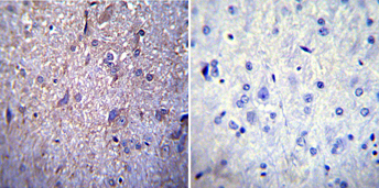
Immunohistochemistry was performed on normal biopsies of de-paraffinized rat brain tissue. To expose target proteins, heat induced antigen retrieval was performed using 10mM sodium citrate (pH 6.0) buffer, microwaved for 8-15 minutes. Following antigen retrieval tissues were blocked in 3% BSA-PBS for 30 minutes at room temperature. Tissues were then probed at a dilution of 1:200 with a mouse monoclonal antibody recognizing Nuclear Pore-O-Linked Glycoprotein or without primary antibody (negative control) overnight at 4°C in a humidified chamber. Tissues were washed extensively with PBST and endogenous peroxidase activity was quenched with a peroxidase suppressor. Detection was performed using a biotin-conjugated secondary antibody and SA-HRP, followed by colorimetric detection using DAB. Tissues were counterstained with hematoxylin and prepped for mounting.Application
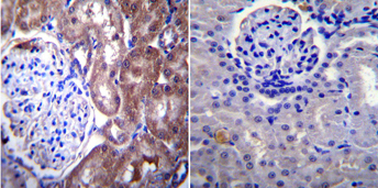
Immunohistochemistry was performed on normal biopsies of de-paraffinized rat kidney tissue. To expose target proteins, heat induced antigen retrieval was performed using 10mM sodium citrate (pH 6.0) buffer, microwaved for 8-15 minutes. Following antigen retrieval tissues were blocked in 3% BSA-PBS for 30 minutes at room temperature. Tissues were then probed at a dilution of 1:200 with a mouse monoclonal antibody recognizing Nuclear Pore-O-Linked Glycoprotein or without primary antibody (negative control) overnight at 4°C in a humidified chamber. Tissues were washed extensively with PBST and endogenous peroxidase activity was quenched with a peroxidase suppressor. Detection was performed using a biotin-conjugated secondary antibody and SA-HRP, followed by colorimetric detection using DAB. Tissues were counterstained with hematoxylin and prepped for mounting.Application
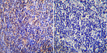
Immunohistochemistry was performed on normal biopsies of de-paraffinized rat lymph node tissue. To expose target proteins, heat induced antigen retrieval was performed using 10mM sodium citrate (pH 6.0) buffer, microwaved for 8-15 minutes. Following antigen retrieval tissues were blocked in 3% BSA-PBS for 30 minutes at room temperature. Tissues were then probed at a dilution of 1:200 with a mouse monoclonal antibody recognizing Nuclear Pore-O-Linked Glycoprotein or without primary antibody (negative control) overnight at 4°C in a humidified chamber. Tissues were washed extensively with PBST and endogenous peroxidase activity was quenched with a peroxidase suppressor. Detection was performed using a biotin-conjugated secondary antibody and SA-HRP, followed by colorimetric detection using DAB. Tissues were counterstained with hematoxylin and prepped for mounting.| Product Name | Nucleoporin p62 antibody |
|---|---|
| Antibody Type | Primary Antibodies |
| Product description | Mouse Monoclonal antibody to Nucleoporin p62Diffusion of metabolites and small non-nuclear molecules as well as active, mediated import of protein and export of protein and RNA through the nuclear envelope occurs through nuclear pore complexes or NPC’s. NPC’s contain up to 100 different polypeptides which have a combined mass of about 125 megadaltons. The channel available for passive transport through the NPC is about 9-10 nm in diameter while carrier mediated changes in the NPC result in a ~25 nm channel used for larger, actively transported molecules. Of the 100 polypeptides, at least 8 of these are O-linked N-acetylglycosamine-modified in mammalian cells. All of the mammalian O-linked glycoproteins contain multiple copies of phenylalanine, glycine dipeptide repeats dispersed throughout part of their sequence. Studies indicate that the NPC O-linked glycoproteins have a direct role in nuclear protein import. |
| Immunogen | Pore complex-lamina fraction purified from rat liver nuclear envelopes. |
| Clonality | Monoclonal |
|---|---|
| Isotype | IgM |
| Clone Number | RL1 |
| Host Species | Mouse |
| Tested Applications | ICC/IFIHCIHC-FIPWB |
| ICC/IF: 1:100: |
|
| Species Reactivity | HumanMouseRatYeastAmphibiansBovineInvertebrates |
| Concentration | 1mg/ml |
| Purification | Affinity purified |
| Gene Synonyms | Nup62 Np62 nucleoporin 62 |
|---|---|
| Target | Nucleoporin p62 |
Application
Immunohistochemistry was performed on normal biopsies of de-paraffinized rat brain tissue. To expose target proteins, heat induced antigen retrieval was performed using 10mM sodium citrate (pH 6.0) buffer, microwaved for 8-15 minutes. Following antigen retrieval tissues were blocked in 3% BSA-PBS for 30 minutes at room temperature. Tissues were then probed at a dilution of 1:200 with a mouse monoclonal antibody recognizing Nuclear Pore-O-Linked Glycoprotein or without primary antibody (negative control) overnight at 4°C in a humidified chamber. Tissues were washed extensively with PBST and endogenous peroxidase activity was quenched with a peroxidase suppressor. Detection was performed using a biotin-conjugated secondary antibody and SA-HRP, followed by colorimetric detection using DAB. Tissues were counterstained with hematoxylin and prepped for mounting.
Application
Immunohistochemistry was performed on normal biopsies of de-paraffinized rat kidney tissue. To expose target proteins, heat induced antigen retrieval was performed using 10mM sodium citrate (pH 6.0) buffer, microwaved for 8-15 minutes. Following antigen retrieval tissues were blocked in 3% BSA-PBS for 30 minutes at room temperature. Tissues were then probed at a dilution of 1:200 with a mouse monoclonal antibody recognizing Nuclear Pore-O-Linked Glycoprotein or without primary antibody (negative control) overnight at 4°C in a humidified chamber. Tissues were washed extensively with PBST and endogenous peroxidase activity was quenched with a peroxidase suppressor. Detection was performed using a biotin-conjugated secondary antibody and SA-HRP, followed by colorimetric detection using DAB. Tissues were counterstained with hematoxylin and prepped for mounting.
Application
Immunohistochemistry was performed on normal biopsies of de-paraffinized rat lymph node tissue. To expose target proteins, heat induced antigen retrieval was performed using 10mM sodium citrate (pH 6.0) buffer, microwaved for 8-15 minutes. Following antigen retrieval tissues were blocked in 3% BSA-PBS for 30 minutes at room temperature. Tissues were then probed at a dilution of 1:200 with a mouse monoclonal antibody recognizing Nuclear Pore-O-Linked Glycoprotein or without primary antibody (negative control) overnight at 4°C in a humidified chamber. Tissues were washed extensively with PBST and endogenous peroxidase activity was quenched with a peroxidase suppressor. Detection was performed using a biotin-conjugated secondary antibody and SA-HRP, followed by colorimetric detection using DAB. Tissues were counterstained with hematoxylin and prepped for mounting.| Application Notes | ICC/IF: 1:100: |
|---|
| Form | Liquid |
|---|---|
| Storage Instructions | Keep as concentrated solution. Aliquot and store at -20ºC or below. Avoid multiple freeze-thaw cycles. |
| Storage Buffer | PBS containing 0.05% sodium azide. |


