Application
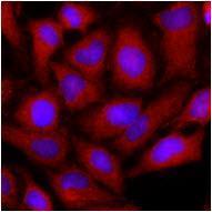
Immunofluorescence of human HeLa cells stained with monoclonal anti-human OAT anitbody (1:500) with Texas Red (Red). Nucleus was stained by Hoechst 33342 (Blue).Application
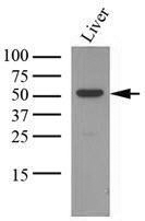
Tissue lysates of mouse liver (35ug) were resolved by SDS-PAGE, transferred to NC membrane and probed with anti-human OAT (1:1000). Proteins were visualized using a goat anti-mouse secondary antibody conjugated to HRP and an ECL detection system.| Product Name | OAT antibody |
|---|---|
| Antibody Type | Primary Antibodies |
| Product description | Mouse monoclonal antibody [AT23A2] to OATOAT is a 49-kDa nucleus-encoded protein imported into mitochondria to give the mature 48-kDa OAT polypeptide. It has been described in humans, animals, insects, plants and microorganisms. Especially OAT have sex-differential expression in the mouse kidney. OAT plays crucial physiological roles in amino acid metabolism. OAT shows a large structural and mechanistic similarity to other enzymes from the subgroup III of aminotransferases, which transfer an amino group from a carbon atom that does not carry a carboxyl function. OAT is essential for nitrogen recycling from arginine but not for the stress-induced proline accumulation. |
| Immunogen | Recombinant human OAT (33-439aa) purified from E.coli |
| Clonality | Monoclonal |
|---|---|
| Isotype | IgG1 |
| Clone Number | AT23A2 |
| Host Species | Mouse |
| Tested Applications | ELISAFACSICC/IFWB |
| FACS:1:200~1:500 ICC/IF:1:200~1:500 WB:1:500~1:2000 Notes:Optimal dilutions/concentrations should be determined by the researcher. |
|
| Species Reactivity | HumanMouse |
| Concentration | 1mg/ml (Please refer to the vial label for the specific concentration) |
| Purification | Affinity purified |
| Gene Synonyms | OATASE Ornithine oxo-acid aminotran ornithine aminotransferase (gyrate atrophy) Ornithineoxoacid aminotransferase. DKFZp781A11155 mitochondrial precursor ornithine aminotransferase precursor EC 2.6.1.13 ornithine aminotransferase precursor EC2.6 |
|---|---|
| Target | OAT |
| Light Chain Type | kappa |
Application
Immunofluorescence of human HeLa cells stained with monoclonal anti-human OAT anitbody (1:500) with Texas Red (Red). Nucleus was stained by Hoechst 33342 (Blue).
Application
Tissue lysates of mouse liver (35ug) were resolved by SDS-PAGE, transferred to NC membrane and probed with anti-human OAT (1:1000). Proteins were visualized using a goat anti-mouse secondary antibody conjugated to HRP and an ECL detection system.| Application Notes | FACS:1:200~1:500 ICC/IF:1:200~1:500 WB:1:500~1:2000 Notes:Optimal dilutions/concentrations should be determined by the researcher. |
|---|
| Form | Liquid |
|---|---|
| Storage Instructions | Can be stored at +4C. For long term storage, aliquot and store at -20C. Avoid repeated freezing and thawing cycles. |
| Storage Buffer | In Phosphate-Buffered Saline (pH 7.4) with 0.1% Sodium Azide |


