Application
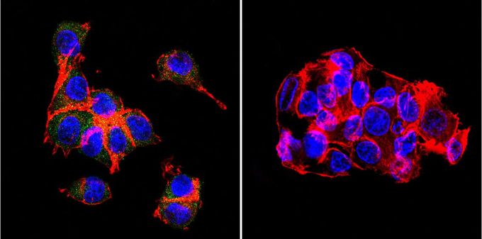
Immunofluorescent analysis of OATP8 in HepG2 cells. Cells were grown on slides and fixed with formaldehyde prior to staining. Cells were probed without (control) or with OATP8 antibody [MDQ] at a dilution of 1:20 overnight at 4 C, washed with PBS and incubated with a proper secondary antibody. OATP8 staining (green), F-Actin staining with Phalloidin (red) and nuclei with DAPI (blue) is shown. Images were taken at 60X magnification.| Product Name | OATP8 antibody |
|---|---|
| Antibody Type | Primary Antibodies |
| Product description | Mouse monoclonal antibody [MDQ] to OATP8This gene encodes a liver-specific member of the organic anion transporter family. The encoded protein is a transmembrane receptor that mediates the sodium-independent uptake of endogenous and xenobiotic compounds and plays a critical role in bile acid and bilirubin transport. Mutations in this gene are a cause of Rotor type hyperbilirubinemia. [provided by RefSeq, Feb 2012] |
| Immunogen | Synthetic peptide corresponding to residues M(1) D Q N Q H L N K T A E A Q P S E N K K T R Y(23) C of human OATP2. |
| Clonality | Monoclonal |
|---|---|
| Isotype | IgG1 |
| Clone Number | MDQ |
| Host Species | Mouse |
| Tested Applications | ICC/IFIHCIHC-FIPWB |
| ICC/IF: 1:20: |
|
| Species Reactivity | HumanMouse |
| Concentration | 1mg/ml |
| Gene Synonyms | LST-3TM13 solute carrier organic anion transporter family member 1B3 SLC21A8 HBLRR LST-2 SLCO1B3 OATP1B3 LST3 OATP-8 |
|---|---|
| Target | OATP8 |
| Tissue Specificity | This antibody also detects the OATP2 isoform. |
Application
Immunofluorescent analysis of OATP8 in HepG2 cells. Cells were grown on slides and fixed with formaldehyde prior to staining. Cells were probed without (control) or with OATP8 antibody [MDQ] at a dilution of 1:20 overnight at 4 C, washed with PBS and incubated with a proper secondary antibody. OATP8 staining (green), F-Actin staining with Phalloidin (red) and nuclei with DAPI (blue) is shown. Images were taken at 60X magnification.| Application Notes | ICC/IF: 1:20: |
|---|
| Form | Liquid |
|---|---|
| Storage Instructions | -20° C, Avoid Freeze/Thaw Cycles |
| Storage Buffer | tissue culture supernatant containing 0.09 % sodium azide |


