Application
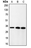
Western blot analysis of CDK5R1 p35 expression in rat brain (A); HeLa (B); A431 (C) whole cell lysates.Application
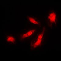
Immunofluorescent analysis of CDK5R1 p35 staining in HeLa cells. Formalin-fixed cells were permeabilized with 0.1% Triton X-100 in TBS for 5-10 minutes and blocked with 3% BSA-PBS for 30 minutes at room temperature. Cells were probed with the primary antibody in 3% BSA-PBS and incubated overnight at 4 C in a humidified chamber. Cells were washed with PBST and incubated with a DyLight 594-conjugated secondary antibody (red) in PBS at room temperature in the dark. DAPI was used to stain the cell nuclei (blue).| Product Name | p35 antibody |
|---|---|
| Antibody Type | Primary Antibodies |
| Product description | Rabbit Polyclonal antibody to p35The protein encoded by this gene (p35) is a neuron-specific activator of cyclin-dependent kinase 5 (CDK5); the activation of CDK5 is required for proper development of the central nervous system. The p35 form of this protein is proteolytically cleaved by calpain, generating a p25 form. The cleavage of p35 into p25 results in relocalization of the protein from the cell periphery to nuclear and perinuclear regions. P25 deregulates CDK5 activity by prolonging its activation and changing its cellular location. The p25 form accumulates in the brain neurons of patients with Alzheimer's disease. This accumulation correlates with an increase in CDK5 kinase activity, and may lead to aberrantly phosphorylated forms of the microtubule-associated protein tau, which contributes to Alzheimer's disease. [provided by RefSeq, Jul 2008] |
| Immunogen | KLH-conjugated synthetic peptide encompassing a sequence within the N-term region of human CDK5R1 p35. The exact sequence is proprietary. |
| Clonality | Polyclonal |
|---|---|
| Isotype | IgG |
| Host Species | Rabbit |
| Tested Applications | ICC/IFWB |
| ICC/IF:1:200~1:500 WB:1:500~1:2000 Notes:Optimal dilutions/concentrations should be determined by the researcher. |
|
| Species Reactivity | HumanMouseRatZebra FishBovine |
| Concentration | 1mg/ml |
| Purification | Affinity purified |
| Gene Synonyms | cyclin-dependent kinase 5 regulatory subunit 1 (p35) p25 p35nck5a CDK5P35 CDK5R NCK5A p23 CDK5R1 |
|---|---|
| Target | p35 |
| Tissue Specificity | Recognizes endogenous levels of CDK5R1 p35 protein. |
Application
Western blot analysis of CDK5R1 p35 expression in rat brain (A); HeLa (B); A431 (C) whole cell lysates.
Application
Immunofluorescent analysis of CDK5R1 p35 staining in HeLa cells. Formalin-fixed cells were permeabilized with 0.1% Triton X-100 in TBS for 5-10 minutes and blocked with 3% BSA-PBS for 30 minutes at room temperature. Cells were probed with the primary antibody in 3% BSA-PBS and incubated overnight at 4 C in a humidified chamber. Cells were washed with PBST and incubated with a DyLight 594-conjugated secondary antibody (red) in PBS at room temperature in the dark. DAPI was used to stain the cell nuclei (blue).| Application Notes | ICC/IF:1:200~1:500 WB:1:500~1:2000 Notes:Optimal dilutions/concentrations should be determined by the researcher. |
|---|
| Form | Liquid |
|---|---|
| Storage Instructions | Shipped at 4 C. Upon delivery aliquot and store at -20 C for one year. Avoid freeze/thaw cycles. |
| Storage Buffer | Liquid in 0.42% Potassium phosphate; 0.87% Sodium chloride; pH 7.3; 30% glycerol; and 0.01% sodium azide. |


