Application
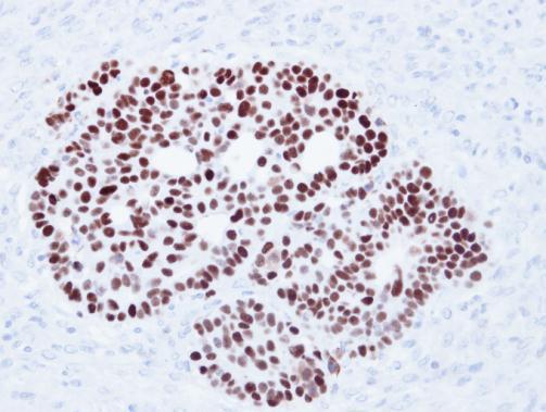
Immunohistochemical analysis of paraffin-embedded Cal27 xenograft, using p53 antibody at 1:500 dilution.Application
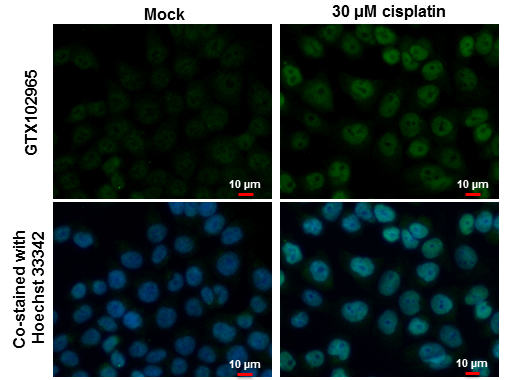
p53 antibody detects p53 protein at nucleus by immunofluorescent analysis. Samples: HCT 116 cells mock (left) and treated with 30 μM Cisplatin for 24 hrs (right) were fixed in 4% paraformaldehyde at RT for 15 min. Green: p53 protein stained by p53 antibody diluted at 1:500. Blue: Hoechst 33342 staining. Scale bar = 10 μm.Application
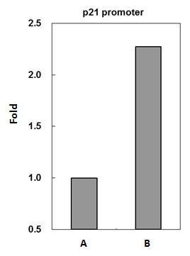
p53 antibody immunoprecipitates p53 protein-DNA complex in ChIP experiments. ChIP Sample: HCT116 whole cell lysate/extract treated with CPT 500nM for 6hr A. 5 μg preimmune rabbit IgG B. 5 μg of p53 antibody The precipitated DNA was detected by PCR with primer set targeting to p21 promoter.Application
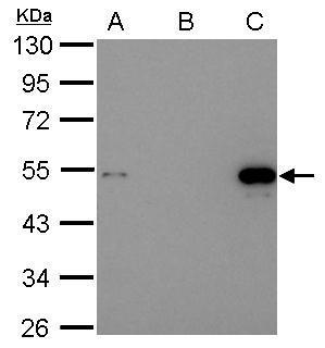
Immunoprecipitation of p53 protein. HCT116 lysates with 30uM cisplatin treatment for 24 hours were subjected to immunoprecipitation using (B) normal rabbit IgG or (C) 2.5 ug of anti-p53 antibody . (A) Input, 20ug of HCT116 lysates. The precipitated protein was detected by diluted at 1:10000. EasyBlot anti-Rabbit IgG Kit was used in Western blot.Application
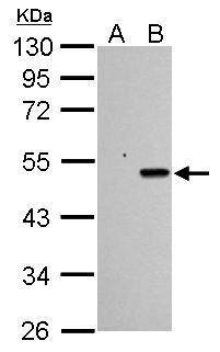
Sample (30 µg of whole cell lysate) A: HCT116 cells with mock treatment for 24 hr B: HCT116 cells with 30 µM cisplatin treatment for 24 hr 10% SDS PAGE diluted at 1:1000 The HRP-conjugated anti-rabbit IgG antibody was used to detect the primary antibody.Application
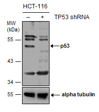
Non-transfected (–) and transfected (+) HCT116 whole cell extracts (30 µg) were separated by 10% SDS-PAGE, and the membrane was blotted with p53 antibody diluted at 1:1000. The HRP-conjugated anti-rabbit IgG antibody was used to detect the primary antibody.Application
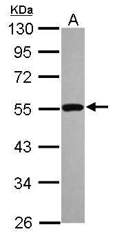
Sample (30 µg of whole cell lysate) A: A431 10% SDS PAGE diluted at 1:1000 The HRP-conjugated anti-rabbit IgG antibody was used to detect the primary antibody.Application
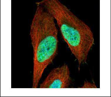
Confocal immunofluorescence analysis (Olympus FV10i) of paraformaldehyde-fixed U2OS, using p53 antibody (Green) at 1:500 dilution. Alpha-tubulin filaments were labeled with (Red) at 1:2000.| Product Name | p53 antibody |
|---|---|
| Antibody Type | Primary Antibodies |
| Product description | Rabbit Polyclonal antibody to p53 (tumor protein p53)This gene encodes tumor protein p53, which responds to diverse cellular stresses to regulate target genes that induce cell cycle arrest, apoptosis, senescence, DNA repair, or changes in metabolism. p53 protein is expressed at low level in normal cells and at a high level in a variety of transformed cell lines, where it's believed to contribute to transformation and malignancy. p53 is a DNA-binding protein containing transcription activation, DNA-binding, and oligomerization domains. It is postulated to bind to a p53-binding site and activate expression of downstream genes that inhibit growth and/or invasion, and thus function as a tumor suppressor. Mutants of p53 that frequently occur in a number of different human cancers fail to bind the consensus DNA binding site, and hence cause the loss of tumor suppressor activity. Alterations of this gene occur not only as somatic mutations in human malignancies, but also as germline mutations in some cancer-prone families with Li-Fraumeni syndrome. Multiple p53 variants due to alternative promoters and multiple alternative splicing have been found. These variants encode distinct isoforms, which can regulate p53 transcriptional activity. [provided by RefSeq] |
| Immunogen | Recombinant protein encompassing a sequence within the center region of human p53. The exact sequence is proprietary. |
| Modification | Acetylated |
| Clonality | Polyclonal |
|---|---|
| Isotype | IgG |
| Host Species | Rabbit |
| Tested Applications | ChIPICC/IFIHC-PIPWB |
| *Optimal dilutions/concentrations should be determined by the researcher.: |
|
| Species Reactivity | HumanMonkeyMouseRat |
| Predicted Reactivity | Rat, Dog, Cat, Pig, Rabbit, Sheep, Rhesus Monkey, Bovine, Guinea pigRat (86%), Dog (91%), Cat (90%), Pig (89%), Rabbit (92%), Sheep (86%), Rhesus Monkey (97%), Bovine (86%), Guinea pig (83%) |
| Concentration | 1mg/ml (Please refer to the vial label for the specific concentration) |
| Purification | Affinity purified |
| Gene Synonyms | FLJ92943 antibody LFS1 antibody P53 antibody TRP53 antibody TP53 antibody cellular tumor antigen p53 antibody antigen NY-CO-13 antibody phosphoprotein p53 antibody p53 tumor suppressor antibody transformation-related protein 53 antibody tumor pro |
|---|---|
| Molecular Weight(MW) | 44 kDa (note) (7) |
| Cellular Localization | Cytoplasm , Nucleus , Endoplasmic reticulum |
Application
Immunohistochemical analysis of paraffin-embedded Cal27 xenograft, using p53 antibody at 1:500 dilution.
Application
p53 antibody detects p53 protein at nucleus by immunofluorescent analysis. Samples: HCT 116 cells mock (left) and treated with 30 μM Cisplatin for 24 hrs (right) were fixed in 4% paraformaldehyde at RT for 15 min. Green: p53 protein stained by p53 antibody diluted at 1:500. Blue: Hoechst 33342 staining. Scale bar = 10 μm.
Application
p53 antibody immunoprecipitates p53 protein-DNA complex in ChIP experiments. ChIP Sample: HCT116 whole cell lysate/extract treated with CPT 500nM for 6hr A. 5 μg preimmune rabbit IgG B. 5 μg of p53 antibody The precipitated DNA was detected by PCR with primer set targeting to p21 promoter.
Application
Immunoprecipitation of p53 protein. HCT116 lysates with 30uM cisplatin treatment for 24 hours were subjected to immunoprecipitation using (B) normal rabbit IgG or (C) 2.5 ug of anti-p53 antibody . (A) Input, 20ug of HCT116 lysates. The precipitated protein was detected by diluted at 1:10000. EasyBlot anti-Rabbit IgG Kit was used in Western blot.
Application
Sample (30 µg of whole cell lysate) A: HCT116 cells with mock treatment for 24 hr B: HCT116 cells with 30 µM cisplatin treatment for 24 hr 10% SDS PAGE diluted at 1:1000 The HRP-conjugated anti-rabbit IgG antibody was used to detect the primary antibody.
Application
Non-transfected (–) and transfected (+) HCT116 whole cell extracts (30 µg) were separated by 10% SDS-PAGE, and the membrane was blotted with p53 antibody diluted at 1:1000. The HRP-conjugated anti-rabbit IgG antibody was used to detect the primary antibody.
Application
Sample (30 µg of whole cell lysate) A: A431 10% SDS PAGE diluted at 1:1000 The HRP-conjugated anti-rabbit IgG antibody was used to detect the primary antibody.
Application
Confocal immunofluorescence analysis (Olympus FV10i) of paraformaldehyde-fixed U2OS, using p53 antibody (Green) at 1:500 dilution. Alpha-tubulin filaments were labeled with (Red) at 1:2000.| Positive Control | 293T , A431 , HCT116 , HCT116 24hr cisplatin 30uM , *HUVEC |
|---|---|
| Application Notes | *Optimal dilutions/concentrations should be determined by the researcher.: |
| Form | Liquid |
|---|---|
| Storage Instructions | Keep as concentrated solution. Aliquot and store at -20ºC or below. Avoid multiple freeze-thaw cycles. |
| Storage Buffer | 1XPBS, 1% BSA, 20% Glycerol (pH7). 0.01% Thimerosal was added as a preservative. |


