Application
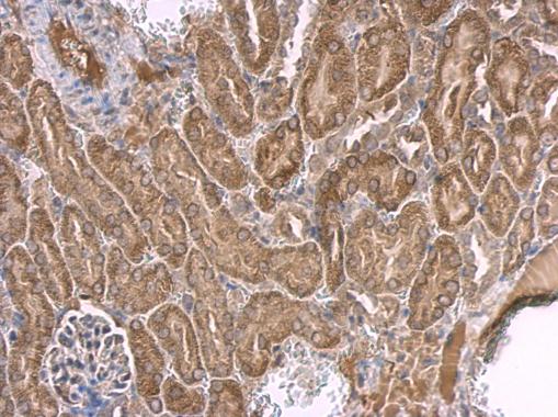
p53 antibody [C2C3], C-term detects p53 protein at cytosol on mouse kidney by immunohistochemical analysis. Sample: Paraffin-embedded mouse kidney. p53 antibody [C2C3], C-term dilution: 1:500.Application
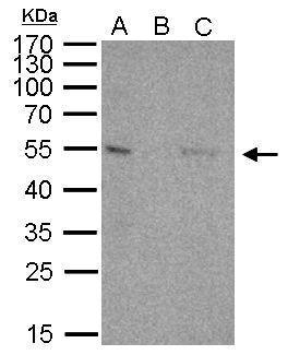
p53 antibody immunoprecipitates p53 protein in IP experiments. IP Sample: 1000 µg 293T whole cell lysate/extract A. 40 μg 293T whole cell lysate/extract B. Control with 2.5 μg of preimmune rabbit IgG C. Immunoprecipitation of p53 protein by 2.5 μg of p53 antibody 12% SDS-PAGE The immunoprecipitated p53 protein was detected by p53 antibody diluted at 1:1000. EasyBlot anti-rabbit IgG was used as a secondary reagent.Application
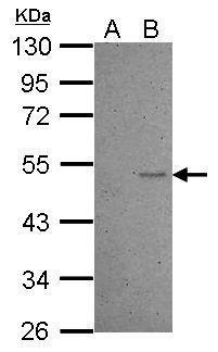
Sample (30 ug of whole cell lysate) A: HCT116 cells with mock treatment for 24hr B: HCT116 cells with 30uM cisplatin treatment for 24hr 10% SDS PAGE diluted at 1:1000WB
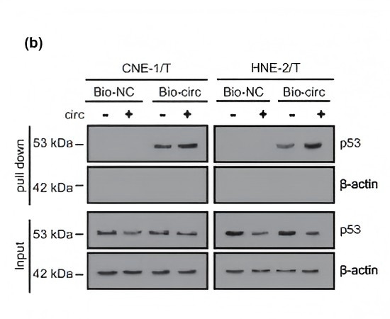
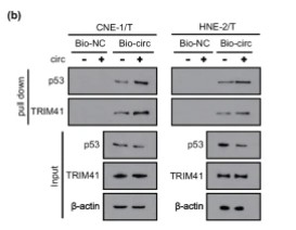
(b) Lysates prepared from HNE-2/T cells and CNE-1/T cells were hybridized with biotinylated circRNA_0067717 probe (bio-circ) and subjected to RNA pull-down assays. Western blot shows that bio-circ pull-down had more p53 protein after circRNA_0067717 overexpression. The bottom panels show the cell lysates’ input. Here, β-Actin functions as the internal control.WB
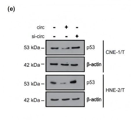
(e) Western blot shows that circRNA_0067717 overexpression resulted in decreased p53 protein level, but the knockdown of circRNA_0067717 increased the p53 level. Statistical analysis of p53 protein relative expression levels in each group.WB
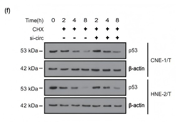
(f) CHX (10 μg/ml) was added to cell culture media and incubated for 0, 4, 8, 12, or 24 h. Immunoblot shows that p53 protein degradation slows after transfection with circRNA_0067717 siRNA. Statistical analysis of the relative p53 protein expression level in HNE-2/T and CNE-1/T cells.WB
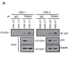
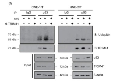
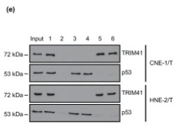
(f) Confirmation of p53 binding to TRIM41 by co-immunoprecipitation (Co-IP) and western blot. The bottom panels show the cell lysates’ input. Here, β-Actin was used as the internal control.WB
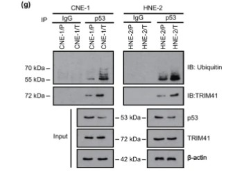
(g) After immunoprecipitation with the p53 antibody, the cell lysates were then analyzed with western blot using anti-ubiquitin and TRIM41 antibodies. Immunoglobulin G served as a negative control.WB
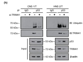
(h) HNE-2/T and CNE-1/T cells transfected with TRIM41 siRNA underwent immunoprecipitation with the anti-p53 antibody and analysis with immunoblot using anti-ubiquitin and TRIM41 antibodies.WB
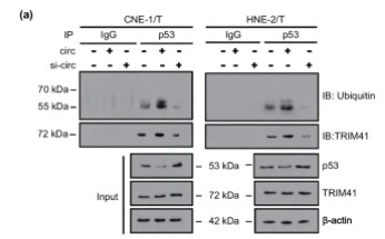
(a) HNE-2/T and CNE-1/T cells underwent transfection with circRNA_0067717 plasmid and circRNA_0067717-specific siRNA, respectively, while the cell lysates underwent immunoprecipitation with the p53 antibody. Both the immunoprecipitate (IP: p53) and cell lysates (Input) were analyzed via western blot using anti-ubiquitin and TRIM41 antibodies.WB
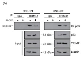
(b) HNE-2/T and CNE-1/T cells transfected with circRNA_0067717 siRNA were subjected to immunoprecipitation with anti-TRIM41 antibody and analyzed with immunoblot using anti-p53. IgG served as a negative control. The bottom panels show the cell lysates’ input.WB
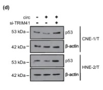
(d) Western blot showing that knockdown of TRIM41 can reverse p53 protein reduction caused by overexpression of circRNA_0067717. Statistical analysis of the relative expression level of p53 protein.WB
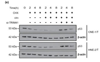
(e) HNE-2/T and CNE-1/T cells were co-transfected with circRNA_0067717 plasmid or TRIM41 siRNA, respectively. 10 μg/ml CHX was added to the cell culture media and incubated for 0, 4, 8, 12, or 24 h. Immunoblot showing that TRIM41 siRNA transfection reversed the rapid degradation of p53 protein caused by circRNA_0067717. Statistical analysis of the relative p53 protein expression levels.WB
(f) Immunoprecipitation followed by ubiquitination experiments showing that circRNA_0067717 overexpression enhanced p53 ubiquitination, while si-TRIM41 attenuated p53 ubiquitination. The bottom panel shows the cell lysate input.WB
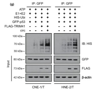
(g) GFP-p53 underwent incubation with 50 nM E1 (UBE1), 500 nM E2 (UbcH5a), 200 μM HIS-ubiquitin, and either FLAG-TRIM41 or circRNA_0067717 overexpression plasmid in 20 μl reaction buffer, then analyzed by immunoprecipitation and immunoblot using the indicated antibodies. The bottom panels depict the input of the cell lysates.WB
(b) RNA pull-down assays showing that overexpression of circRNA_0067717 can pull down more TRIM41 and p53. The bottom panels show the cell lysate inputs.WB
(e) Western blot detected the expression levels of TRIM41 and p53 in circRNA_0067717 different probes.WB
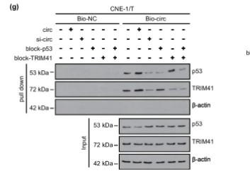
(g) RNA pull-down experiments depicting block-p53’s attenuation of p53 the binding to circRNA_0067717 but does not affect the binding of TRIM41 to circRNA_0067717, similar to block-TRIM41 in CNE-1/T cells.WB
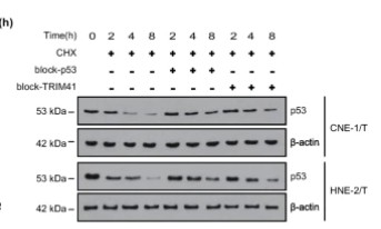
(h) HNE-2/T and CNE-1/T cells were co-transfected with block-p53 and block-TRIM41, respectively 10 μg/ml of CHX was added to cell culture media. Western blot showed that block-p53 and block-TRIM41 enhance the protein stability of p53.WB
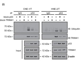
(i) Immunoprecipitation followed by ubiquitination experiments showing that block-p53 and block-TRIM41 can reduce the ubiquitination level of p53 and promote its protein expression. The bottom panels show the cell lysate inputs. β-actin served as an internal control.WB
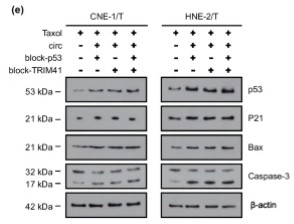
(e) Western blot showing that block-p53 or block-TRIM41 can promote p53, p21, Bax, and caspase-3 protein expression. Statistical analysis of the relative expression level of each protein.| Product Name | p53 antibody , C-term |
|---|---|
| Antibody Type | Primary Antibodies |
| Product description | Rabbit Polyclonal antibody to p53 (tumor protein p53)This gene encodes tumor protein p53, which responds to diverse cellular stresses to regulate target genes that induce cell cycle arrest, apoptosis, senescence, DNA repair, or changes in metabolism. p53 protein is expressed at low level in normal cells and at a high level in a variety of transformed cell lines, where it's believed to contribute to transformation and malignancy. p53 is a DNA-binding protein containing transcription activation, DNA-binding, and oligomerization domains. It is postulated to bind to a p53-binding site and activate expression of downstream genes that inhibit growth and/or invasion, and thus function as a tumor suppressor. Mutants of p53 that frequently occur in a number of different human cancers fail to bind the consensus DNA binding site, and hence cause the loss of tumor suppressor activity. Alterations of this gene occur not only as somatic mutations in human malignancies, but also as germline mutations in some cancer-prone families with Li-Fraumeni syndrome. Multiple p53 variants due to alternative promoters and multiple alternative splicing have been found. These variants encode distinct isoforms, which can regulate p53 transcriptional activity. [provided by RefSeq] |
| Immunogen | Carrier-protein conjugated synthetic peptide encompassing a sequence within the C-terminus region of human p53. The exact sequence is proprietary. |
| Clonality | Polyclonal |
|---|---|
| Isotype | IgG |
| Host Species | Rabbit |
| Tested Applications | IHC-PIPWB |
| *Optimal dilutions/concentrations should be determined by the researcher.
: |
|
| Species Reactivity | HumanMouse |
| Predicted Reactivity | Sheep, Rhesus MonkeySheep (100%), Rhesus Monkey (100%) |
| Concentration | 1mg/ |
| Purification | Affinity purified |
| Gene Synonyms | FLJ92943 antibody LFS1 antibody P53 antibody TRP53 antibody TP53 antibody cellular tumor antigen p53 antibody antigen NY-CO-13 antibody phosphoprotein p53 antibody p53 tumor suppressor antibody transformation-related protein 53 antibody tumor pro |
|---|---|
| Molecular Weight(MW) | 44 kDa (note) (8) |
| Cellular Localization | Cytoplasm , Nucleus , Endoplasmic reticulum |
Application
p53 antibody [C2C3], C-term detects p53 protein at cytosol on mouse kidney by immunohistochemical analysis. Sample: Paraffin-embedded mouse kidney. p53 antibody [C2C3], C-term dilution: 1:500.
Application
p53 antibody immunoprecipitates p53 protein in IP experiments. IP Sample: 1000 µg 293T whole cell lysate/extract A. 40 μg 293T whole cell lysate/extract B. Control with 2.5 μg of preimmune rabbit IgG C. Immunoprecipitation of p53 protein by 2.5 μg of p53 antibody 12% SDS-PAGE The immunoprecipitated p53 protein was detected by p53 antibody diluted at 1:1000. EasyBlot anti-rabbit IgG was used as a secondary reagent.
Application
Sample (30 ug of whole cell lysate) A: HCT116 cells with mock treatment for 24hr B: HCT116 cells with 30uM cisplatin treatment for 24hr 10% SDS PAGE diluted at 1:1000
WB
(b) Lysates prepared from HNE-2/T cells and CNE-1/T cells were hybridized with biotinylated circRNA_0067717 probe (bio-circ) and subjected to RNA pull-down assays. Western blot shows that bio-circ pull-down had more p53 protein after circRNA_0067717 overexpression. The bottom panels show the cell lysates’ input. Here, β-Actin functions as the internal control.
WB
(e) Western blot shows that circRNA_0067717 overexpression resulted in decreased p53 protein level, but the knockdown of circRNA_0067717 increased the p53 level. Statistical analysis of p53 protein relative expression levels in each group.
WB
(f) CHX (10 μg/ml) was added to cell culture media and incubated for 0, 4, 8, 12, or 24 h. Immunoblot shows that p53 protein degradation slows after transfection with circRNA_0067717 siRNA. Statistical analysis of the relative p53 protein expression level in HNE-2/T and CNE-1/T cells.
WB
(f) Confirmation of p53 binding to TRIM41 by co-immunoprecipitation (Co-IP) and western blot. The bottom panels show the cell lysates’ input. Here, β-Actin was used as the internal control.
WB
(g) After immunoprecipitation with the p53 antibody, the cell lysates were then analyzed with western blot using anti-ubiquitin and TRIM41 antibodies. Immunoglobulin G served as a negative control.
WB
(h) HNE-2/T and CNE-1/T cells transfected with TRIM41 siRNA underwent immunoprecipitation with the anti-p53 antibody and analysis with immunoblot using anti-ubiquitin and TRIM41 antibodies.
WB
(a) HNE-2/T and CNE-1/T cells underwent transfection with circRNA_0067717 plasmid and circRNA_0067717-specific siRNA, respectively, while the cell lysates underwent immunoprecipitation with the p53 antibody. Both the immunoprecipitate (IP: p53) and cell lysates (Input) were analyzed via western blot using anti-ubiquitin and TRIM41 antibodies.
WB
(b) HNE-2/T and CNE-1/T cells transfected with circRNA_0067717 siRNA were subjected to immunoprecipitation with anti-TRIM41 antibody and analyzed with immunoblot using anti-p53. IgG served as a negative control. The bottom panels show the cell lysates’ input.
WB
(d) Western blot showing that knockdown of TRIM41 can reverse p53 protein reduction caused by overexpression of circRNA_0067717. Statistical analysis of the relative expression level of p53 protein.
WB
(e) HNE-2/T and CNE-1/T cells were co-transfected with circRNA_0067717 plasmid or TRIM41 siRNA, respectively. 10 μg/ml CHX was added to the cell culture media and incubated for 0, 4, 8, 12, or 24 h. Immunoblot showing that TRIM41 siRNA transfection reversed the rapid degradation of p53 protein caused by circRNA_0067717. Statistical analysis of the relative p53 protein expression levels.
WB
(f) Immunoprecipitation followed by ubiquitination experiments showing that circRNA_0067717 overexpression enhanced p53 ubiquitination, while si-TRIM41 attenuated p53 ubiquitination. The bottom panel shows the cell lysate input.
WB
(g) GFP-p53 underwent incubation with 50 nM E1 (UBE1), 500 nM E2 (UbcH5a), 200 μM HIS-ubiquitin, and either FLAG-TRIM41 or circRNA_0067717 overexpression plasmid in 20 μl reaction buffer, then analyzed by immunoprecipitation and immunoblot using the indicated antibodies. The bottom panels depict the input of the cell lysates.
WB
(b) RNA pull-down assays showing that overexpression of circRNA_0067717 can pull down more TRIM41 and p53. The bottom panels show the cell lysate inputs.
WB
(e) Western blot detected the expression levels of TRIM41 and p53 in circRNA_0067717 different probes.
WB
(g) RNA pull-down experiments depicting block-p53’s attenuation of p53 the binding to circRNA_0067717 but does not affect the binding of TRIM41 to circRNA_0067717, similar to block-TRIM41 in CNE-1/T cells.
WB
(h) HNE-2/T and CNE-1/T cells were co-transfected with block-p53 and block-TRIM41, respectively 10 μg/ml of CHX was added to cell culture media. Western blot showed that block-p53 and block-TRIM41 enhance the protein stability of p53.
WB
(i) Immunoprecipitation followed by ubiquitination experiments showing that block-p53 and block-TRIM41 can reduce the ubiquitination level of p53 and promote its protein expression. The bottom panels show the cell lysate inputs. β-actin served as an internal control.
WB
(e) Western blot showing that block-p53 or block-TRIM41 can promote p53, p21, Bax, and caspase-3 protein expression. Statistical analysis of the relative expression level of each protein.| Positive Control | HCT116 24hr cisplatin 30uM |
|---|---|
| Application Notes | *Optimal dilutions/concentrations should be determined by the researcher.
: |
| Form | Liquid |
|---|---|
| Storage Instructions | Keep as concentrated solution. Aliquot and store at -20ºC or below. Avoid multiple freeze-thaw cycles. |
| Storage Buffer | 1XPBS, 1% BSA, 20% Glycerol (pH7). 0.01% Thimerosal was added as a preservative. |


