Application
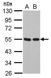
Sample (30 µg of whole cell lysate) A: 293T B: A431 10% SDS PAGE diluted at 1:10000 The HRP-conjugated anti-mouse IgG antibody was used to detect the primary antibody.Application
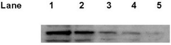
The image shows a Western blot for , testing the threshold detection of the p53 antibody. The lanes are as follows:. Lane 1 - 20ug of A549 lysate, lane 2 - 10ug of A549 lysate, lane 3 - 5ug of A549 lysate, lane 4 - 2.5ug of A549 lysate, laneApplication
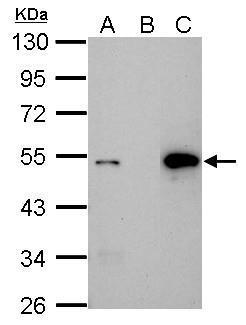
Immunoprecipitation of p53 protein. HCT116 lysates with 30uM cisplatin treatment for 24 hours were subjected to immunoprecipitation using (B) normal mouse IgG or (C) 2.5 ug of anti-p53 antibody . (A) Input, 20ug of HCT116 lysate. The precipitated protein was detected by diluted at 1:10000. EasyBlot anti-Mouse IgG Kit was used in Western blot.Application
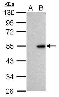
Sample (30 µg of whole cell lysate) A: HCT116 cells with mock treatment for 24 hr B: HCT116 cells with 30 µM cisplatin treatment for 24 hr 10% SDS PAGE diluted at 1:1000 The HRP-conjugated anti-mouse IgG antibody was used to detect the primary antibody.Application
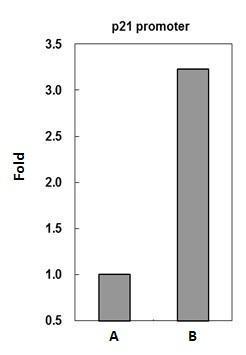
p53 antibody immunoprecipitates p53 protein-DNA complex in ChIP experiments. ChIP Sample: HCT116 whole cell lysate/extract treated with CPT 500nM for 6hr A. 5 μg preimmune mouse IgG B. 5 μg of p53 antibody The precipitated DNA was detected by PCR with primer set targeting to p21 promoter.Application
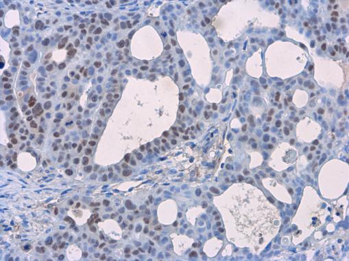
p53 antibody [Pab1801] detects p53 protein at nucleus in NCl-H1299 xenograft by immunohistochemical analysis. Sample: Paraffin-embedded NCl-H1299 xenograft. p53 antibody [Pab1801] diluted at 1:200.| Product Name | p53 antibody |
|---|---|
| Antibody Type | Primary Antibodies |
| Product description | Mouse monoclonal antibody to human p53PAb1801 specifically recognizes human p53 protein, a tumor suppressor gene that is mutated in a wide variety of sporadic and familial cancers. p53, a nuclear protein, mediates cell cycle arrest in response to DNA damage and therefore is important in the maintenance of genomic stability. Recent results indicate that an enhanced phosphorylation of p53 by ATM in response to DNA damage on SER15 is a mutation which reduces the ability of p53 to arrest cell growth. |
| Immunogen | Gel-purified fusion protein containing the coding region of human p53 expressed in E. coli. |
| Clonality | Monoclonal |
|---|---|
| Isotype | IgG1 |
| Clone Number | Pab1801 |
| Host Species | Mouse |
| Tested Applications | ELISAFACSIHC-FIHC-PIPRIAWBChIP |
| *Optimal dilutions/concentrations should be determined by the researcher.
: |
|
| Species Reactivity | Human |
| Concentration | 1mg/ml (Please refer to the vial label for the specific concentration) |
| Purification | Affinity purified |
| Gene Synonyms | Pab1801 Pab 1801 Antigen NY CO 13 Cellular tumor antigen p53 Phosphoprotein p53 TP 53 TP53 Transformation Related Protein p53 TRP 53 TRP53 Tumor protein p53 Li Fraumeni syndrome Tumor suppressor p53 Tumour Protein p53 |
|---|---|
| Target | TP53 |
| Cellular Localization | Cytoplasmic and Nuclear |
| Light Chain Type | kappa |
Application
Sample (30 µg of whole cell lysate) A: 293T B: A431 10% SDS PAGE diluted at 1:10000 The HRP-conjugated anti-mouse IgG antibody was used to detect the primary antibody.
Application
The image shows a Western blot for , testing the threshold detection of the p53 antibody. The lanes are as follows:. Lane 1 - 20ug of A549 lysate, lane 2 - 10ug of A549 lysate, lane 3 - 5ug of A549 lysate, lane 4 - 2.5ug of A549 lysate, lane
Application
Immunoprecipitation of p53 protein. HCT116 lysates with 30uM cisplatin treatment for 24 hours were subjected to immunoprecipitation using (B) normal mouse IgG or (C) 2.5 ug of anti-p53 antibody . (A) Input, 20ug of HCT116 lysate. The precipitated protein was detected by diluted at 1:10000. EasyBlot anti-Mouse IgG Kit was used in Western blot.
Application
Sample (30 µg of whole cell lysate) A: HCT116 cells with mock treatment for 24 hr B: HCT116 cells with 30 µM cisplatin treatment for 24 hr 10% SDS PAGE diluted at 1:1000 The HRP-conjugated anti-mouse IgG antibody was used to detect the primary antibody.
Application
p53 antibody immunoprecipitates p53 protein-DNA complex in ChIP experiments. ChIP Sample: HCT116 whole cell lysate/extract treated with CPT 500nM for 6hr A. 5 μg preimmune mouse IgG B. 5 μg of p53 antibody The precipitated DNA was detected by PCR with primer set targeting to p21 promoter.
Application
p53 antibody [Pab1801] detects p53 protein at nucleus in NCl-H1299 xenograft by immunohistochemical analysis. Sample: Paraffin-embedded NCl-H1299 xenograft. p53 antibody [Pab1801] diluted at 1:200.| Positive Control | HBL100 , HCT116 , 293T , A431 |
|---|---|
| Application Notes | *Optimal dilutions/concentrations should be determined by the researcher.
: |
| Form | Liquid |
|---|---|
| Storage Instructions | Keep as concentrated solution. Store at 4°C short term. For extended storage aliquot and store at -20°C or below. Avoid freeze-thaw cycles. |
| Storage Buffer | Phosphate-buffered saline, pH 7.4, containing no preservatives |


