Application
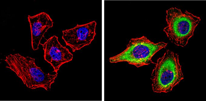
Immunofluorescent analysis of PASK (green) in HeLa cells (right) compared to a negative control without primary antibody (left). Formalin-fixed cells were permeabilized with 0.1% Triton X-100 in TBS for 5-10 minutes and blocked with 3% BSA-PBS for 30 minutes at room temperature. Cells were probed with PASK antibody [mAB6] in 3% BSA-PBS at a dilution of 1:100 and incubated overnight at 4°C in a humidified chamber. Cells were washed with PBST and incubated with a proper secondary antibody in PBS at room temperature in the dark. F-actin (red) was stained with a flourescent red phalloidin and nuclei (blue) were stained with Hoechst or DAPI. Images were taken at a magnification of 60x.| Product Name | PASK antibody |
|---|---|
| Antibody Type | Primary Antibodies |
| Product description | Mouse monoclonal antibody [mAB6] to PASKThis gene encodes a member of the serine/threonine kinase family that contains two PAS domains. Expression of this gene is regulated by glucose, and the encoded protein plays a role in the regulation of insulin gene expression. Downregulation of this gene may play a role in type 2 diabetes. Alternatively spliced transcript variants encoding multiple isoforms have been observed for this gene. [provided by RefSeq, Nov 2011] |
| Immunogen | Recombinant, GST tagged, PAS fusion protein expressed in E. coli. |
| Clonality | Monoclonal |
|---|---|
| Isotype | IgG2 |
| Clone Number | mAB6 |
| Host Species | Mouse |
| Tested Applications | ICC/IFIHCWB |
| ICC/IF: 1:10 - 1:200: |
|
| Species Reactivity | HumanRatPrimate |
| Concentration | 1mg/ml (Please refer to the vial label for the specific concentration) |
| Purification | Protein A |
| Gene Synonyms | PASKIN STK37 PAS domain containing serine/threonine kinase |
|---|---|
| Target | PASK |
Application
Immunofluorescent analysis of PASK (green) in HeLa cells (right) compared to a negative control without primary antibody (left). Formalin-fixed cells were permeabilized with 0.1% Triton X-100 in TBS for 5-10 minutes and blocked with 3% BSA-PBS for 30 minutes at room temperature. Cells were probed with PASK antibody [mAB6] in 3% BSA-PBS at a dilution of 1:100 and incubated overnight at 4°C in a humidified chamber. Cells were washed with PBST and incubated with a proper secondary antibody in PBS at room temperature in the dark. F-actin (red) was stained with a flourescent red phalloidin and nuclei (blue) were stained with Hoechst or DAPI. Images were taken at a magnification of 60x.| Application Notes | ICC/IF: 1:10 - 1:200: |
|---|
| Form | Liquid |
|---|---|
| Storage Instructions | -20° C, Avoid Freeze/Thaw Cycles |
| Storage Buffer | PBS containing 1 mg/ml BSA and 0.05 % sodium azide |


