Application
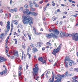
Immunohistochemical analysis of paraffin-embedded human placenta tissues using PEG10 mouse mAbApplication
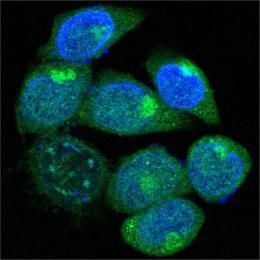
Confocal immunofluorescence analysis of methanol-fixed HepG2 cells using anti-PEG10 monoclonal antioby (green), showing cytoplasmic localization. Blue: DRAQ5 fluorescent DNA dye.Application
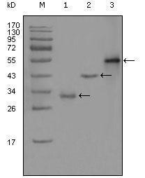
Western blot analysis using PEG10 monoclonal antibody against truncated Trx-PEG10 recombinant protein(1),truncated GST-PEG10(aa1-120) recombinant protein(2) and full-length PEG10(aa1-325)-hIgGFc transfected CHO-K1 cell lysate(3). .| Product Name | PEG10 antibody |
|---|---|
| Antibody Type | Primary Antibodies |
| Product description | Mouse monoclonal antibody [1B1C4] to PEG10This is a paternally expressed imprinted gene that encodes transcripts containing two overlapping open reading frames (ORFs), RF1 and RF1/RF2, as well as retroviral-like slippage and pseudoknot elements, which can induce a -1 nucleotide frame-shift. ORF1 encodes a shorter isoform with a CCHC-type zinc finger motif containing a sequence characteristic of gag proteins of most retroviruses and some retrotransposons. The longer isoform is the result of -1 translational frame-shifting leading to translation of a gag/pol-like protein combining RF1 and RF2. It contains the active-site consensus sequence of the protease domain of pol proteins. Additional isoforms resulting from alternatively spliced transcript variants, as well as from use of upstream non-AUG (CUG) start codon, have been reported for this gene. Increased expression of this gene is associated with hepatocellular carcinomas. [provided by RefSeq, May 2010] |
| Immunogen | The immunogen corresponding to a region within amino acids 1 and 120 of PEG10 expressed in E. Coli. |
| Clonality | Monoclonal |
|---|---|
| Isotype | IgG1 |
| Clone Number | 1B1C4 |
| Host Species | Mouse |
| Tested Applications | ELISAICC/IFIHC-PWB |
| ELISA: 1/10000.: |
|
| Species Reactivity | Human |
| Concentration | 1mg/ml |
| Gene Synonyms | EDR RGAG3 MART2 HB-1 MEF3L MAR2 |
|---|---|
| Target | PEG10 |
| Cellular Localization | Detected predominantly in the cytoplasm of breast and prostate carcinomas, in hepatocellular carcinoma (HCC) and B-cell chronic lymphocytic leukemia (B-CLL) cells and in the HepG2 cell line. Colocalized with ALK1. |
Application
Immunohistochemical analysis of paraffin-embedded human placenta tissues using PEG10 mouse mAb
Application
Confocal immunofluorescence analysis of methanol-fixed HepG2 cells using anti-PEG10 monoclonal antioby (green), showing cytoplasmic localization. Blue: DRAQ5 fluorescent DNA dye.
Application
Western blot analysis using PEG10 monoclonal antibody against truncated Trx-PEG10 recombinant protein(1),truncated GST-PEG10(aa1-120) recombinant protein(2) and full-length PEG10(aa1-325)-hIgGFc transfected CHO-K1 cell lysate(3). .| Application Notes | ELISA: 1/10000.: |
|---|
| Form | Liquid |
|---|---|
| Storage Instructions | Store at 4℃, for long term storage, store at -20℃. |
| Storage Buffer | Ascitic fluid containing 0.03% sodium azide. |


