Application
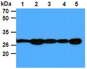
The Cell lysates (40ug) were resolved by SDS-PAGE, transferred to PVDF membrane and probed with anti-human PGAM1 antibody (1:1000). Proteins were visualized using a goat anti-mouse secondary antibody conjugated to HRP and an ECL detection system. Lane 1: 293T cell lysate Lane. 2: Jurkat cell lysate Lane. 3: Raji cell lysate Lane. 4: A431 cell lysate Lane. 5: HeLa cell lysate.Application
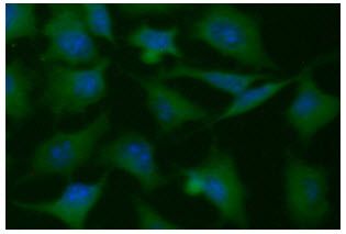
ICC/IF analysis of PGAM1 in HeLa cells line, stained with DAPI (Blue) for nucleus staining and monoclonal anti-human PGAM1 antibody (1:100) with goat anti-mouse IgG-Alexa fluor 488 conjugate (Green).Application
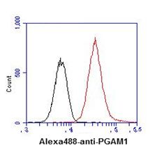
Flow cytometry analysis of PGAM1 in HeLa cell line, staining at 2-5ug for 1x10*6 cells (red line). The secondary antibody used goat anti-mouse IgG Alexa fluor 488 conjugate. Isotype control antibody was mouse IgG (black line).| Product Name | PGAM1 antibody |
|---|---|
| Antibody Type | Primary Antibodies |
| Product description | Mouse Monoclonal antibody [AT1G4] to PGAM1Phosphoglyceric acid mutase (EC 2.7.5.3) is widely distributed in mammalian tissues where it catalyzes the reversible reaction of 3-phosphoglycerate (3-PGA) to 2-phosphoglycerate (2-PGA) in the glycolytic pathway (summary by Chen et al., 1974 [PubMed 4811757]).[supplied by OMIM, Nov 2010] |
| Immunogen | Recombinant human PGAM1 (1-254aa) purified from E. coli |
| Clonality | Monoclonal |
|---|---|
| Isotype | IgG2a |
| Clone Number | AT1G4 |
| Host Species | Mouse |
| Tested Applications | ELISAFACSICC/IFWB |
| WB: 1:1000. ICC/IF: 1:100: |
|
| Species Reactivity | Human |
| Concentration | 1mg/ml (Please refer to the vial label for the specific concentration) |
| Purification | Affinity purified |
| Gene Synonyms | phosphoglycerate mutase 1 (brain) PGAM-B PGAMA HEL-S-35 |
|---|---|
| Target | PGAM1 |
| Light Chain Type | kappa |
Application
The Cell lysates (40ug) were resolved by SDS-PAGE, transferred to PVDF membrane and probed with anti-human PGAM1 antibody (1:1000). Proteins were visualized using a goat anti-mouse secondary antibody conjugated to HRP and an ECL detection system. Lane 1: 293T cell lysate Lane. 2: Jurkat cell lysate Lane. 3: Raji cell lysate Lane. 4: A431 cell lysate Lane. 5: HeLa cell lysate.
Application
ICC/IF analysis of PGAM1 in HeLa cells line, stained with DAPI (Blue) for nucleus staining and monoclonal anti-human PGAM1 antibody (1:100) with goat anti-mouse IgG-Alexa fluor 488 conjugate (Green).
Application
Flow cytometry analysis of PGAM1 in HeLa cell line, staining at 2-5ug for 1x10*6 cells (red line). The secondary antibody used goat anti-mouse IgG Alexa fluor 488 conjugate. Isotype control antibody was mouse IgG (black line).| Application Notes | WB: 1:1000. ICC/IF: 1:100: |
|---|
| Form | Liquid |
|---|---|
| Storage Instructions | Can be stored at +4C. For long term storage, aliquot and store at -20C. Avoid repeated freezing and thawing cycles. |
| Storage Buffer | Liquid. In Phosphate-Buffered Saline (pH 7.4) with 0.02% Sodium Azide, 10% Glycerol. |


