Application
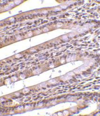
Immunohistochemical staining of PHAP in human small intestine with PHAP antibody at 10μg/ml.Application
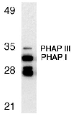
Western blot analysis of PHAP expression in Raji cell lysate with PHAP antibody at 1μg/ml. The two bands below PHAP I might be differently spliced isoforms of PHAP.| Product Name | PHAP antibody |
|---|---|
| Antibody Type | Primary Antibodies |
| Product description | Rabbit polyclonal to PHAPApoptosis is related to many diseases and development. Caspase 9 plays a central role in cell death induced by a variety of apoptosis activators. Cytochrome c, after released from mitochondria, binds to Apaf 1, which forms an apoptosome that in turn binds to and activate procaspase 9. Activated caspase 9 cleaves and activates the effector caspases (caspase 3, 6 and 7), which are responsible for the proteolytic cleavage of many key proteins in apoptosis. The tumor suppressor putative HLA-DR-associated proteins (PHAPs) were recently identified as important regulators of mitochondrion apoptosis. PHAP appears to facilitate apoptosome-mediated caspase 9 activation and to stimulate the mitochondrial apoptotic pathway. PHAP was also shown to oppose both Ras- and Myc-medicated cell transformation. |
| Immunogen | Synthetic peptide (Human). |
| Clonality | Polyclonal |
|---|---|
| Isotype | IgG |
| Host Species | Rabbit |
| Tested Applications | IHCWB |
| WB: Use at a concentration of 0.5 - 2.0 μg/ml. Detects bands of approximately 35 and 32 kDa corresponding to PHAPIII and PHAPI respectively. Two cross reacting bands are also observed below 32 kDa, which may be differentially spliced forms of PHAPI. Optimal dilutions/concentrations should be determined by the end user.: |
|
| Species Reactivity | HumanMouseRat |
| Concentration | 1mg/ml (Please refer to the vial label for the specific concentration) |
| Purification | Affinity purified |
| Gene Synonyms | C15orf1 I1PP2A LANP MAPM MGC119787 MGC150373 PHAP1 PHAPI PP32 |
|---|---|
| Target | PHAP |
| Tissue Specificity | This antibody cross-reacts with both PHAP I and PHAP III. |
| Cellular Localization | Cytoplasmic and Nuclear |
Application
Immunohistochemical staining of PHAP in human small intestine with PHAP antibody at 10μg/ml.
Application
Western blot analysis of PHAP expression in Raji cell lysate with PHAP antibody at 1μg/ml. The two bands below PHAP I might be differently spliced isoforms of PHAP.| Positive Control | Raji whole cell lysate |
|---|---|
| Application Notes | WB: Use at a concentration of 0.5 - 2.0 μg/ml. Detects bands of approximately 35 and 32 kDa corresponding to PHAPIII and PHAPI respectively. Two cross reacting bands are also observed below 32 kDa, which may be differentially spliced forms of PHAPI. Optimal dilutions/concentrations should be determined by the end user.: |
| Form | Liquid |
|---|---|
| Storage Instructions | For short-term storage, store at 4° C. For long-term storage, aliquot and store at -20ºC or below. Avoid multiple freeze-thaw cycles. |
| Storage Buffer | Preservative: 0.02% Sodium Azide; Constituents: PBS |


