Application
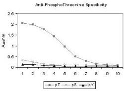
ELISA results of Mab anti-phosphothreonine antibody tested against BSA conjugates of pT, pY and pS. Each well was coated with 0.1µg of conjugate. The starting dilution of antibody was 1:1000 and each point on the X-axis represents a 2-fold dilution. HRP conjugated Gt-a-Mouse IgG (H&L) antibody and TMB substrate were used for detection.Application
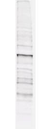
Mab anti-phosphothreonine antibody (clone 18F6, ) is shown to detect threonine phosphorylation of proteins in a lysate from A431 cells stimulated with EGF. Separation is achieved under reducing conditions using a pre-cast 4-20% iGel from Gradipore, Inc. A 1:1,000 dilution of Mab anti-phosphothreonine is used for 30 min followed by detection using 1:2,500 dilution Goat-a-Mouse IgG [H&L] secondary antibody and visualization using the Odyssey® Infrared Imaging System developed by LI-COR.| Product Name | Phosphothreonine antibody |
|---|---|
| Antibody Type | Primary Antibodies |
| Product description | Mouse Monoclonal antibody [18F6] to PhosphothreoninePhosphorylation of threonine residues is associated with many growth factors and oncogene protein kinases, and is important for cell signaling in activation, proliferation and differentiation. |
| Immunogen | Phosphothreonine antibody was produced after repeated immunizations of balb/c mice with phosphothreonine conjugated KLH. |
| Clonality | Monoclonal |
|---|---|
| Isotype | IgG1 |
| Clone Number | 18F6 |
| Host Species | Mouse |
| Tested Applications | ELISAWB |
| *Optimal dilutions/concentrations should be determined by the researcher.
: |
|
| Concentration | 1mg/ml |
| Purification | Affinity purified |
| Target | Phosphothreonine |
|---|
Application
ELISA results of Mab anti-phosphothreonine antibody tested against BSA conjugates of pT, pY and pS. Each well was coated with 0.1µg of conjugate. The starting dilution of antibody was 1:1000 and each point on the X-axis represents a 2-fold dilution. HRP conjugated Gt-a-Mouse IgG (H&L) antibody and TMB substrate were used for detection.
Application
Mab anti-phosphothreonine antibody (clone 18F6, ) is shown to detect threonine phosphorylation of proteins in a lysate from A431 cells stimulated with EGF. Separation is achieved under reducing conditions using a pre-cast 4-20% iGel from Gradipore, Inc. A 1:1,000 dilution of Mab anti-phosphothreonine is used for 30 min followed by detection using 1:2,500 dilution Goat-a-Mouse IgG [H&L] secondary antibody and visualization using the Odyssey® Infrared Imaging System developed by LI-COR.| Application Notes | *Optimal dilutions/concentrations should be determined by the researcher.
: |
|---|
| Form | Liquid |
|---|---|
| Storage Instructions | Store vial at -20° C prior to opening. Aliquot contents and freeze at -20° C or below for extended storage. Avoid cycles of freezing and thawing. |
| Storage Buffer | Ascites with 0.01% (w/v) Sodium Azide |


