Application
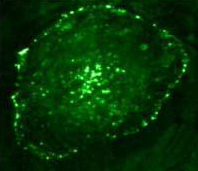
Immunofluorescence with anti-Phosphotyrosine Monoclonal Antibody [IG2]Application
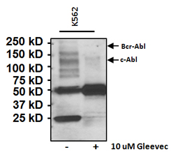
Immunoprecipitation of Phosphotyrosine (p-Tyr) was performed on K562 cell lysates from untreated cells (left lane) or from cells treated with 10uM of the tyrosine kinase inhibitor Gleevec (right lane) for 24 hours. Antigen-antibody complexes were formed by incubating 500ug of whole cell lysate with 2ug of a p-Tyr monoclonal antibody overnight on a rocking platform at 4°C. The immune complexes were captured on 50ul Protein A/G Agarose, washed extensively, and eluted with Lane Marker Reducing Sample Buffer. Samples were resolved on a 4-20% Tris-HCl polyacrylamide gel, transferred to a PVDF membrane, and blocked with 5% BSA/TBST for at least 1 hour. The membrane was probed with a c-Abl polyclonal antibody at a dilution of 1:1000 overnight rotating at 4°C, washed in TBST, and probed with goat anti-rabbit IgG-HRP secondary antibody) at a dilution of 1:20,000 for at least 1 hour. Chemiluminescent detection was performedApplication
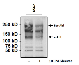
Western blot analysis of tyrosine phosphorylated c-Abl and Bcr-Abl was performed following immunoprecipitation of K562 cell lysates using a c-Abl antibody, from untreated cells (left lane) or cells treated with 10uM of the tyrosine kinase inhibitor Gleevec (right lane) for 24 hours. The captured immune complexes were resolved on a 4-20% Tris-HCl polyacrylamide gel, transferred to a PVDF membrane, and blocked with 5% BSA/TBST for at least 1 hour. The membrane was probed with a Phosphotyrosine monoclonal antibody at a dilution of 1:250 overnight rotating at 4°C, washed in TBST, and probed with a goat anti-mouse IgG-HRP secondary antibody at a dilution of 1:20,000 for at least 1 hour. Chemiluminescent detection was performed| Product Name | Phosphotyrosine antibody |
|---|---|
| Antibody Type | Primary Antibodies |
| Product description | Mouse monoclonal [IG2] to PhosphotyrosineProtein kinases like platelet-derived growth factor and epidermal growth factor have the ability to phosphorylate tyrosine residues of other proteins like GTPase activating protein. Protein kinases play a prominent role in the regulation of normal growth as well as tumor growth. A number of retroviruses encode proteins with tyrosine kinase activity and infection with such a virus is associated with a 10-20 fold increase in phosphotyrosine on the virally encoded protein as well as on other cellular proteins. This antibody, from clone 1G2, is capable of specifically binding to proteins which contain phosphorylated tyrosine residues, but will not react with any other phosphorylated amino acids, proteins or nuecleotides, and is excellent for the study of growth factor receptors, oncogenes, and signal transduction. |
| Immunogen | O-phospho-L-tyrosine and O-phospho-DL-tyramine. |
| Clonality | Monoclonal |
|---|---|
| Isotype | IgG1 |
| Clone Number | IG2 |
| Host Species | Mouse |
| Tested Applications | ELISAFACSICC/IFIHCIPWB |
| ICC/IF: 1:50 - 1:200: |
|
| Species Reactivity | HumanMouseRatChemical |
| Concentration | 1mg/ml (Please refer to the vial label for the specific concentration) |
| Purification | Affinity purified |
| Target | Phosphotyrosine |
|---|---|
| Tissue Specificity | detects phosphotyrosine from most species and tissues. will not react with any other phosphorylated amino acids, proteins or nucleotides. |
Application
Immunofluorescence with anti-Phosphotyrosine Monoclonal Antibody [IG2]
Application
Immunoprecipitation of Phosphotyrosine (p-Tyr) was performed on K562 cell lysates from untreated cells (left lane) or from cells treated with 10uM of the tyrosine kinase inhibitor Gleevec (right lane) for 24 hours. Antigen-antibody complexes were formed by incubating 500ug of whole cell lysate with 2ug of a p-Tyr monoclonal antibody overnight on a rocking platform at 4°C. The immune complexes were captured on 50ul Protein A/G Agarose, washed extensively, and eluted with Lane Marker Reducing Sample Buffer. Samples were resolved on a 4-20% Tris-HCl polyacrylamide gel, transferred to a PVDF membrane, and blocked with 5% BSA/TBST for at least 1 hour. The membrane was probed with a c-Abl polyclonal antibody at a dilution of 1:1000 overnight rotating at 4°C, washed in TBST, and probed with goat anti-rabbit IgG-HRP secondary antibody) at a dilution of 1:20,000 for at least 1 hour. Chemiluminescent detection was performed
Application
Western blot analysis of tyrosine phosphorylated c-Abl and Bcr-Abl was performed following immunoprecipitation of K562 cell lysates using a c-Abl antibody, from untreated cells (left lane) or cells treated with 10uM of the tyrosine kinase inhibitor Gleevec (right lane) for 24 hours. The captured immune complexes were resolved on a 4-20% Tris-HCl polyacrylamide gel, transferred to a PVDF membrane, and blocked with 5% BSA/TBST for at least 1 hour. The membrane was probed with a Phosphotyrosine monoclonal antibody at a dilution of 1:250 overnight rotating at 4°C, washed in TBST, and probed with a goat anti-mouse IgG-HRP secondary antibody at a dilution of 1:20,000 for at least 1 hour. Chemiluminescent detection was performed| Positive Control | HeLa cells |
|---|---|
| Application Notes | ICC/IF: 1:50 - 1:200: |
| Form | Liquid |
|---|---|
| Storage Instructions | Store at 4ºC for short-term storage. Aliquot and store at -20ºC for long-term. Avoid repeated freezing and thawing. |
| Storage Buffer | Preservative: 0.05% Sodium Azide; Constituents: 40% Glycerol, PBS |


