Application
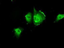
Anti-PIM2 mouse monoclonal antibody immunofluorescent staining of COS7 cells transiently transfected with PIM2Application
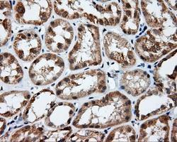
Immunohistochemical staining of paraffin-embedded Kidney tissue using anti-RC201933 mouse monoclonal antibody. ( Dilution 1:50)Application
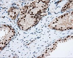
Immunohistochemical staining of paraffin-embedded prostate tissue using anti-RC201933 mouse monoclonal antibody. ( Dilution 1:50)Application
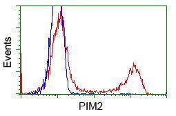
HEK293T cells transfected with either pCMV6-ENTRY PIM2Application
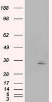
HEK293T cells were transfected with the pCMV6-ENTRY control (Left lane) or pCMV6-ENTRY PIM2 (Right lane) cDNA for 48 hrs and lysed. Equivalent amounts of cell lysates (5 ug per lane) were separated by SDS-PAGE and immunoblotted with anti-PIM2.Application
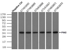
Immunoprecipitation(IP) of PIM2 by using TrueMab monoclonal anti-PIM2 antibodies (Negative control: IP without adding anti-PIM2 antibody). For each experiment, 500ul of DDK tagged PIM2 overexpression lysates (at 1:5 dilution with HEK293T lysate), 2ug of anti-PIM2 antibody and 20ul (0.1mg) of goat anti-mouse conjugated magnetic beads were mixed and incubated overnight. After extensive wash to remove any non-specific binding, the immuno-precipitated products were analyzed with rabbit anti-DDK polyclonal antibody.| Product Name | PIM2 antibody [8B4] |
|---|---|
| Antibody Type | Primary Antibodies |
| Product description | Mouse monoclonal antibody [8B4] to PIM2Promotes cell survival in response to a variety of proliferative signals via positive regulation of the I-kappaB kinase/NF-kappaB cascade | this process requires phosphorylation of MAP3K8/COT. Prevents apoptosis induced by growth factor withdrawal via inhibition of caspase-3 activation, and via phosphorylation of pro-apoptotic proteins. Inhibits BAD-induced cell death via phosphorylation of BAD. PIM2-mediated cell survival is glucose-dependent but independent of several AKT regulators such as PI3K, HSP-90 and TOR, indicating that PIM2 and PI3K/AKT/TOR function via distinct pathways. Involved in the positive regulation of chondrocyte survival and autophagy in the epiphyseal growth plate. |
| Immunogen | Full length human recombinant protein of human PIM2 (NP_006866) produced in HEK293T cell. |
| Clonality | Monoclonal |
|---|---|
| Isotype | IgG2a |
| Clone Number | 8B4 |
| Host Species | Mouse |
| Tested Applications | FACSICC/IFIHCIHC-PIPWB |
| IHC 1:50 | |
| Species Reactivity | Human |
| Concentration | 1mg/ml |
| Purification | Affinity purified |
| Target | PIM2 |
|---|---|
| Tissue Specificity | is specific for human PIM2 (NP_006866). |
Application
Anti-PIM2 mouse monoclonal antibody immunofluorescent staining of COS7 cells transiently transfected with PIM2
Application
Immunohistochemical staining of paraffin-embedded Kidney tissue using anti-RC201933 mouse monoclonal antibody. ( Dilution 1:50)
Application
Immunohistochemical staining of paraffin-embedded prostate tissue using anti-RC201933 mouse monoclonal antibody. ( Dilution 1:50)
Application
HEK293T cells transfected with either pCMV6-ENTRY PIM2
Application
HEK293T cells were transfected with the pCMV6-ENTRY control (Left lane) or pCMV6-ENTRY PIM2 (Right lane) cDNA for 48 hrs and lysed. Equivalent amounts of cell lysates (5 ug per lane) were separated by SDS-PAGE and immunoblotted with anti-PIM2.
Application
Immunoprecipitation(IP) of PIM2 by using TrueMab monoclonal anti-PIM2 antibodies (Negative control: IP without adding anti-PIM2 antibody). For each experiment, 500ul of DDK tagged PIM2 overexpression lysates (at 1:5 dilution with HEK293T lysate), 2ug of anti-PIM2 antibody and 20ul (0.1mg) of goat anti-mouse conjugated magnetic beads were mixed and incubated overnight. After extensive wash to remove any non-specific binding, the immuno-precipitated products were analyzed with rabbit anti-DDK polyclonal antibody.| Application Notes | IHC 1:50 |
|---|
| Form | Liquid |
|---|---|
| Storage Instructions | For short-term storage, store at 4°C or aliquot into working amounts and store at -20°C. For long-term storage, store at -70°C (aliquotted). Avoid repeated freeze-thaw cycles. |
| Storage Buffer | PBS (PH 7.3) containing 1% BSA, 50% glycerol and 0.02% sodium azide. |


