Application
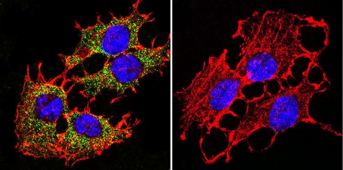
Immunofluorescent analysis of PMCA ATPase in C6 cells. PMCA ATPase staining (green), F-Actin staining with Phalloidin (red) and nuclei with DAPI (blue) is shown. Cells were grown on slides and fixed with formaldehyde prior to staining. Cells were probed without (control) or with PMCA ATPase antibody [5F10] at a dilution of 1:200 over night at 4?C, washed with PBS and incubated with a proper secondary antibody. Images were taken at 60X magnification.| Product Name | PMCA ATPase antibody |
|---|---|
| Antibody Type | Primary Antibodies |
| Product description | Mouse monoclonal antibody [5F10] to PMCA ATPaseThe protein encoded by this gene belongs to the family of P-type primary ion transport ATPases characterized by the formation of an aspartyl phosphate intermediate during the reaction cycle. These enzymes remove bivalent calcium ions from eukaryotic cells against very large concentration gradients and play a critical role in intracellular calcium homeostasis. The mammalian plasma membrane calcium ATPase isoforms are encoded by at least four separate genes and the diversity of these enzymes is further increased by alternative splicing of transcripts. The expression of different isoforms and splice variants is regulated in a developmental, tissue- and cell type-specific manner, suggesting that these pumps are functionally adapted to the physiological needs of particular cells and tissues. This gene encodes the plasma membrane calcium ATPase isoform 1. Alternatively spliced transcript variants encoding different isoforms have been identified. [provided by RefSeq, Jul 2008] |
| Immunogen | Purified human erythrocyte calcium ATPase. |
| Clonality | Monoclonal |
|---|---|
| Isotype | IgG2a |
| Clone Number | 5F10 |
| Host Species | Mouse |
| Tested Applications | BlockingELISAFACSICC/IFIHCIHC-FIHC-PIPWB |
| ICC/IF: 1:100 - 1:500: |
|
| Species Reactivity | HumanMouseChickenDogPigRatAmphibiansCatHamsterPrimateRabbitSheepBovineVertebratesAvian |
| Concentration | 1mg/ml |
| Gene Synonyms | ATPase Ca++ transporting plasma membrane 1 PMCA1kb ATP2B1 PMCA1 |
|---|---|
| Target | PMCA ATPase |
| Tissue Specificity | This antibody detects all four known isoforms of the PMCA ATPase. |
Application
Immunofluorescent analysis of PMCA ATPase in C6 cells. PMCA ATPase staining (green), F-Actin staining with Phalloidin (red) and nuclei with DAPI (blue) is shown. Cells were grown on slides and fixed with formaldehyde prior to staining. Cells were probed without (control) or with PMCA ATPase antibody [5F10] at a dilution of 1:200 over night at 4?C, washed with PBS and incubated with a proper secondary antibody. Images were taken at 60X magnification.| Application Notes | ICC/IF: 1:100 - 1:500: |
|---|
| Form | Liquid |
|---|---|
| Storage Instructions | -20° C, Avoid Freeze/Thaw Cycles |
| Storage Buffer | Ascites containing 0.05 % sodium azide |


