Application
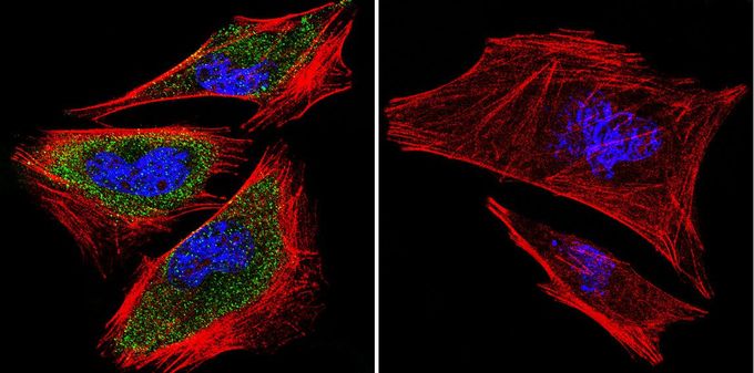
Immunofluorescent analysis of PRC1 in A2058 cells. PRC1 staining (green), F-Actin staining with Phalloidin (red) and nuclei with DAPI (blue) is shown. Cells were grown on slides and fixed with formaldehyde prior to staining. Cells were probed without (control) or with PRC1 antibody [16F2] at a dilution of 1:100 over night at 4°C, washed with PBS and incubated with a proper secondary antibody. Images were taken at 60X magnification.| Product Name | PRC1 antibody |
|---|---|
| Antibody Type | Primary Antibodies |
| Product description | Mouse monoclonal antibody [16F2] to PRC1This gene encodes a protein that is involved in cytokinesis. The protein is present at high levels during the S and G2/M phases of mitosis but its levels drop dramatically when the cell exits mitosis and enters the G1 phase. It is located in the nucleus during interphase, becomes associated with mitotic spindles in a highly dynamic manner during mitosis, and localizes to the cell mid-body during cytokinesis. This protein has been shown to be a substrate of several cyclin-dependent kinases (CDKs). It is necessary for polarizing parallel microtubules and concentrating the factors responsible for contractile ring assembly. Alternative splicing results in multiple transcript variants. [provided by RefSeq, Jun 2012] |
| Immunogen | Recombinant PRC1 protein. |
| Clonality | Monoclonal |
|---|---|
| Isotype | IgG2 |
| Clone Number | 16F2 |
| Host Species | Mouse |
| Tested Applications | ICC/IFIPWB |
| ICC/IF: 1:100 Optimal dilution / concentration should be determined by the end user.: |
|
| Species Reactivity | HumanMouseRat |
| Concentration | 1mg/ml (Please refer to the vial label for the specific concentration) |
| Purification | Protein G |
| Gene Synonyms | ASE1 protein regulator of cytokinesis 1 |
|---|---|
| Target | PRC1 |
Application
Immunofluorescent analysis of PRC1 in A2058 cells. PRC1 staining (green), F-Actin staining with Phalloidin (red) and nuclei with DAPI (blue) is shown. Cells were grown on slides and fixed with formaldehyde prior to staining. Cells were probed without (control) or with PRC1 antibody [16F2] at a dilution of 1:100 over night at 4°C, washed with PBS and incubated with a proper secondary antibody. Images were taken at 60X magnification.| Application Notes | ICC/IF: 1:100 Optimal dilution / concentration should be determined by the end user.: |
|---|
| Form | Liquid |
|---|---|
| Storage Instructions | -20° C, Avoid Freeze/Thaw Cycles |
| Storage Buffer | PBS containing 1 mg/ml BSA and 0.05 % sodium azide |


