Application
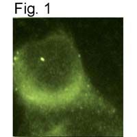
Immunocytochemical staining of Presenilin-1 in mouse fibroblasts.Application
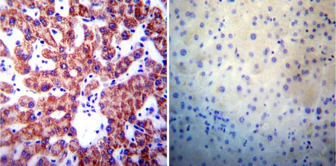
Immunohistochemistry was performed on normal biopsies of deparaffinized human liver tissue. To expose target proteins, heat induced antigen retrieval was performed using 10mM sodium citrate (pH6.0) buffer, microwaved for 8-15 minutes. Following antigen retrieval tissues were blocked in 3% BSA-PBS for 30 minutes at room temperature. Tissues were then probed at a dilution of 1:200 with or without Presenilin-1 antibody [APS 18] overnight at 4°C in a humidified chamber. Tissues were washed extensively with PBST and endogenous peroxidase activity was quenched with a peroxidase suppressor. Detection was performed using a biotin-conjugated secondary antibody and SA-HRP, followed by colorimetric detection using DAB. Tissues were counterstained with hematoxylin and prepped for mounting.Application
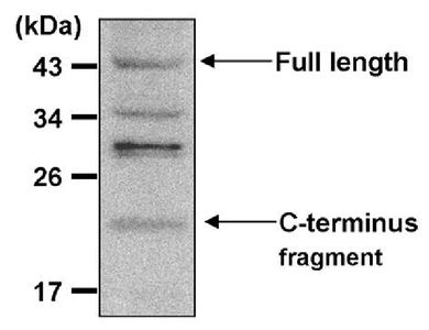
Western blot analysis of Presenilin-1 in 20ug of THP-1 cell lysate. Proteins were transferred to a PVDF membrane and blocked with 5% non-fat milk in TBST for 1 hour at room temperature. The membrane was probed with Presenilin-1 antibody [APS 18] at a dilution of 1:250 overnight at 4oC, washed in TBST, and probed with a HRP-conjugated secondary antibody at a dilution of 1:40,000 for 1 hour at room temperature. Chemiluminescent detection was performed using ECL substrate.| Product Name | Presenilin 1 antibody |
|---|---|
| Antibody Type | Primary Antibodies |
| Product description | Mouse monoclonal antibody [APS 18] to Presenilin 1Alzheimer's disease (AD) patients with an inherited form of the disease carry mutations in the presenilin proteins (PSEN1; PSEN2) or in the amyloid precursor protein (APP). These disease-linked mutations result in increased production of the longer form of amyloid-beta (main component of amyloid deposits found in AD brains). Presenilins are postulated to regulate APP processing through their effects on gamma-secretase, an enzyme that cleaves APP. Also, it is thought that the presenilins are involved in the cleavage of the Notch receptor, such that they either directly regulate gamma-secretase activity or themselves are protease enzymes. Several alternatively spliced transcript variants encoding different isoforms have been identified for this gene, the full-length nature of only some have been determined. [provided by RefSeq, Aug 2008] |
| Immunogen | Synthetic peptide corresponding to residues C S(313) K Y N A E S T E R E S Q D T V A E N D D G(334) of humen Presenilin 1. |
| Clonality | Monoclonal |
|---|---|
| Isotype | IgG1 |
| Clone Number | APS 18 |
| Host Species | Mouse |
| Tested Applications | ELISAICC/IFIHCIHC-PIPWB |
| ICC/IF: 1:50 - 1:200: |
|
| Species Reactivity | HumanMouseRatPrimate |
| Concentration | 1mg/ml (Please refer to the vial label for the specific concentration) |
| Purification | Protein G |
| Gene Synonyms | PS1 FAD PS-1 AD3 PSEN1 S182 presenilin 1 |
|---|---|
| Target | Presenilin 1 |
| Tissue Specificity | No cross-reactivity is seen with Presenilin-2. |
Application
Immunocytochemical staining of Presenilin-1 in mouse fibroblasts.
Application
Immunohistochemistry was performed on normal biopsies of deparaffinized human liver tissue. To expose target proteins, heat induced antigen retrieval was performed using 10mM sodium citrate (pH6.0) buffer, microwaved for 8-15 minutes. Following antigen retrieval tissues were blocked in 3% BSA-PBS for 30 minutes at room temperature. Tissues were then probed at a dilution of 1:200 with or without Presenilin-1 antibody [APS 18] overnight at 4°C in a humidified chamber. Tissues were washed extensively with PBST and endogenous peroxidase activity was quenched with a peroxidase suppressor. Detection was performed using a biotin-conjugated secondary antibody and SA-HRP, followed by colorimetric detection using DAB. Tissues were counterstained with hematoxylin and prepped for mounting.
Application
Western blot analysis of Presenilin-1 in 20ug of THP-1 cell lysate. Proteins were transferred to a PVDF membrane and blocked with 5% non-fat milk in TBST for 1 hour at room temperature. The membrane was probed with Presenilin-1 antibody [APS 18] at a dilution of 1:250 overnight at 4oC, washed in TBST, and probed with a HRP-conjugated secondary antibody at a dilution of 1:40,000 for 1 hour at room temperature. Chemiluminescent detection was performed using ECL substrate.| Application Notes | ICC/IF: 1:50 - 1:200: |
|---|
| Form | Liquid |
|---|---|
| Storage Instructions | -20° C, Avoid Freeze/Thaw Cycles |
| Storage Buffer | PBS containing 0.05 % sodium azide and 1 mg/ml BSA |


