Application
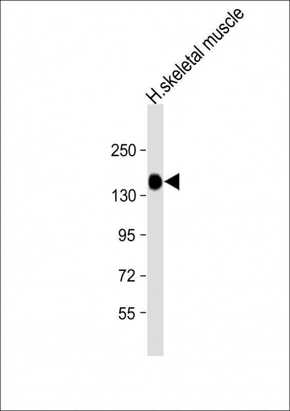
Western Blot at 1:8000 dilution + human skeletal muscle lysate Lysates/proteins at 20 ug per lane.Application
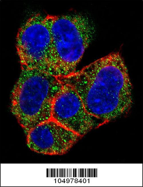
Confocal immunofluorescent analysis of AGL Antibody with HepG2 cell followed by Alexa Fluor 488-conjugated goat anti-rabbit lgG (green). Actin filaments have been labeled with Alexa Fluor 555 phalloidin (red).DAPI was used to stain the cell nuclear (blue).Application
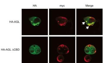
Expression of myc-GS causes wild type but not the ÃCBD mutant of AGL to aggregate around the PAS-stain-positive inclusions. HepG2 cells were transfected with either HA-tagged wild-type AGL (HA-AGL) or HA-AGL ÃCBD. Cells were fixed in formalin and processed for IF using anti-HA (green) and anti-myc (red) antibodies. White arrows indicate colocalization of HA-AGL and myc-GS.Application
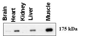
Western blot using anti-AGL antibody at 1:1000 dilution. A total of 20 ug of lysates was loaded for each tissue. Data courtesy of Dr. Alan Cheng, Department of Internal Medicine, Life Sciences Institute, University of Michigan Medical Center, Ann Arbor, Michigan.| Product Name | AGL Antibody |
|---|---|
| Antibody Type | Primary Antibodies |
| Antigen Alias | Glycogen debranching enzyme, Glycogen debrancher, 4-alpha-glucanotransferase, Oligo-1,4-1,4-glucantransferase, Amylo-alpha-1,6-glucosidase, Amylo-1,6-glucosidase, Dextrin 6-alpha-D-glucosidase, AGL, GDE |
| Product description | AGL is a glycogen debrancher enzyme which is involved in glycogen degradation. This enzyme has two independent catalytic activities which occur at different sites on the protein: a 4-alpha-glucotransferase activity and a amylo-1,6-glucosidase activity. Mutations in the AGL gene are associated with glycogen storage disease although a wide range of enzymatic and clinical variability occurs which may be due to tissue-specific alternative splicing.1) Horinishi, A., et al., J. Hum. Genet. 47(2):55-59 (2002). |
| Immunogen | This AGL antibody is generated from rabbits immunized with a KLH conjugated synthetic peptide between 1479-1510 amino acids from the C-terminal region of human AGL. |
| Clonality | Polyclonal |
|---|---|
| Isotype | Ig |
| Host Species | Rabbit |
| Tested Applications | IFWB |
| For WB starting dilution is: 1:1000 For IF starting dilution is: 1:10~50: |
|
| Species Reactivity | Human |
| Concentration | 1mg/ml |
| Gene Symbol | AGL |
|---|---|
| Alternative Names | Glycogen debranching enzyme Glycogen debrancher 4-alpha-glucanotransferase Oligo-1 4-1 4-glucantransferase Amylo-alpha-1 6-glucosidase Amylo-1 6-glucosidase Dextrin 6-alpha-D-glucosidase AGL GDE |
| Molecular Weight(MW) | 175 kDa |
Application
Western Blot at 1:8000 dilution + human skeletal muscle lysate Lysates/proteins at 20 ug per lane.
Application
Confocal immunofluorescent analysis of AGL Antibody with HepG2 cell followed by Alexa Fluor 488-conjugated goat anti-rabbit lgG (green). Actin filaments have been labeled with Alexa Fluor 555 phalloidin (red).DAPI was used to stain the cell nuclear (blue).
Application
Expression of myc-GS causes wild type but not the ÃCBD mutant of AGL to aggregate around the PAS-stain-positive inclusions. HepG2 cells were transfected with either HA-tagged wild-type AGL (HA-AGL) or HA-AGL ÃCBD. Cells were fixed in formalin and processed for IF using anti-HA (green) and anti-myc (red) antibodies. White arrows indicate colocalization of HA-AGL and myc-GS.
Application
Western blot using anti-AGL antibody at 1:1000 dilution. A total of 20 ug of lysates was loaded for each tissue. Data courtesy of Dr. Alan Cheng, Department of Internal Medicine, Life Sciences Institute, University of Michigan Medical Center, Ann Arbor, Michigan.| Application Notes | For WB starting dilution is: 1:1000 For IF starting dilution is: 1:10~50: |
|---|
| Form | Liquid |
|---|---|
| Storage Instructions | Store at 4˚C for three months and -20˚C, stable for up to one year. As with all antibodies care should be taken to avoid repeated freeze thaw cycles. Antibodies should not be exposed to prolonged high temperatures. |
| Storage Buffer | Supplied in PBS with 0.09% (W/V) sodium azide. |


