Application
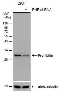
Non-transfected (–) and transfected (+) 293T whole cell extracts (30 µg) were separated by 12% SDS-PAGE, and the membrane was blotted with Prohibitin antibody diluted at 1:2000. The HRP-conjugated anti-rabbit IgG antibody was used to detect the primary antibody.Application
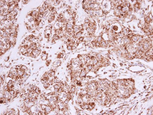
Prohibitin antibody detects Prohibitin protein at mitochondria in human breast carcinoma by immunohistochemical analysis. Sample: Paraffin-embedded human breast carcinoma. Prohibitin antibody diluted at 1:500.Application
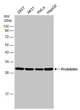
Various whole cell extracts (30 µg) were separated by 12% SDS-PAGE, and the membrane was blotted with Prohibitin antibody diluted at 1:1000.Application
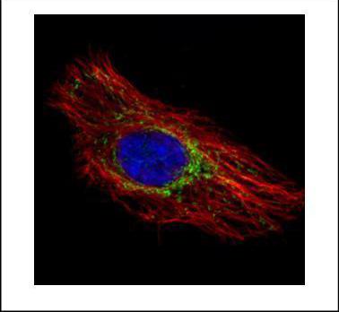
Confocal immunofluorescence analysis (Olympus FV10i) of methanol-fixed HeLa, using Prohibitin antibody (Green) at 1:500 dilution. Alpha-tubulin filaments were labeled with (Red) at 1:2000.Application
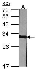
Sample (30 µg of whole cell lysate) A:NIH-3T3 12% SDS PAGE diluted at 1:1000 The HRP-conjugated anti-rabbit IgG antibody was used to detect the primary antibody.Application
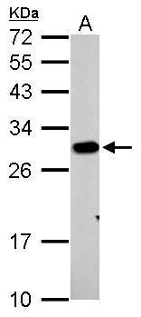
Prohibitin antibody detects PHB protein by western blot analysis. A. 30 µg PC-12 whole cell lysate/extract 12% SDS-PAGE Prohibitin antibody dilution: 1:1000 The HRP-conjugated anti-rabbit IgG antibody was used to detect the primary antibody.Application
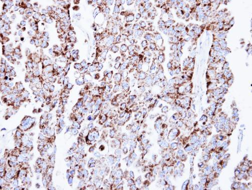
Immunohistochemical analysis of paraffin-embedded OVCAR3 xenograft, using Prohibitin antibody at 1:500 dilution.Application
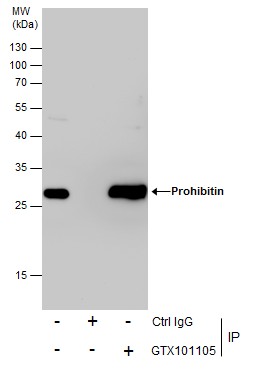
Immunoprecipitation of Prohibitin protein from 293T whole cell extracts using 5 μg of Prohibitin antibody . Western blot analysis was performed using Prohibitin antibody . EasyBlot anti-Rabbit IgG was used as a secondary reagent.Application
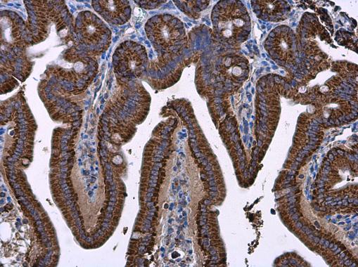
Prohibitin antibody detects Prohibitin protein at cytoplasm in mouse intestine by immunohistochemical analysis. Sample: Paraffin-embedded mouse intestine. Prohibitin antibody diluted at 1:500.Application
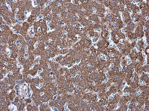
Prohibitin antibody detects Prohibitin protein at cytoplasm in rat liver by immunohistochemical analysis. Sample: Paraffin-embedded rat liver. Prohibitin antibody diluted at 1:500.Application
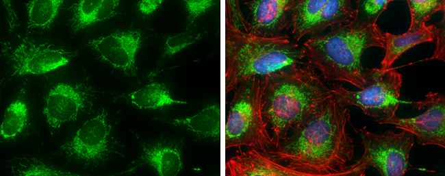
Prohibitin antibody detects Prohibitin protein at mitochondria by immunofluorescent analysis. Sample: HeLa cells were fixed in 4% paraformaldehyde at RT for 15 min. Green: Prohibitin protein stained by Prohibitin antibody diluted at 1:500. Red: Phalloidin, a cytoskeleton marker, diluted at 1:100. Blue: Hoechst 33342 staining.| Product Name | Prohibitin antibody |
|---|---|
| Antibody Type | Primary Antibodies |
| Product description | Rabbit Polyclonal antibody to Prohibitin (prohibitin)Prohibitin is an evolutionarily conserved gene that is ubiquitously expressed. It is thought to be a negative regulator of cell proliferation and may be a tumor suppressor. Mutations in PHB have been linked to sporadic breast cancer. Prohibitin is expressed as two transcripts with varying lengths of 3' untranslated region. The longer transcript is present at higher levels in proliferating tissues and cells, suggesting that this longer 3' untranslated region may function as a trans-acting regulatory RNA. [provided by RefSeq] |
| Immunogen | Recombinant protein encompassing a sequence within the center region of human Prohibitin. The exact sequence is proprietary. |
| Clonality | Polyclonal |
|---|---|
| Isotype | IgG |
| Host Species | Rabbit |
| Tested Applications | ICC/IFIHC-PIPWB |
| *Optimal dilutions/concentrations should be determined by the researcher.
: |
|
| Species Reactivity | HumanMouseRat |
| Predicted Reactivity | Zebrafish, Xenopus laevis, Dog, Cat, Chicken, Rhesus Monkey, Chimpanzee, BovineZebrafish (93%), Xenopus laevis (92%), Dog (100%), Cat (100%), Chicken (95%), Rhesus Monkey (100%), Chimpanzee (100%), Bovine (100%) |
| Concentration | 1mg/ml |
| Purification | Affinity purified |
| Gene Synonyms | PHB1 antibody PHB antibody prohibitin antibody |
|---|---|
| Molecular Weight(MW) | 30 kDa (note) (3) |
| Cellular Localization | Mitochondrion inner membrane |
Application
Non-transfected (–) and transfected (+) 293T whole cell extracts (30 µg) were separated by 12% SDS-PAGE, and the membrane was blotted with Prohibitin antibody diluted at 1:2000. The HRP-conjugated anti-rabbit IgG antibody was used to detect the primary antibody.
Application
Prohibitin antibody detects Prohibitin protein at mitochondria in human breast carcinoma by immunohistochemical analysis. Sample: Paraffin-embedded human breast carcinoma. Prohibitin antibody diluted at 1:500.
Application
Various whole cell extracts (30 µg) were separated by 12% SDS-PAGE, and the membrane was blotted with Prohibitin antibody diluted at 1:1000.
Application
Confocal immunofluorescence analysis (Olympus FV10i) of methanol-fixed HeLa, using Prohibitin antibody (Green) at 1:500 dilution. Alpha-tubulin filaments were labeled with (Red) at 1:2000.
Application
Sample (30 µg of whole cell lysate) A:NIH-3T3 12% SDS PAGE diluted at 1:1000 The HRP-conjugated anti-rabbit IgG antibody was used to detect the primary antibody.
Application
Prohibitin antibody detects PHB protein by western blot analysis. A. 30 µg PC-12 whole cell lysate/extract 12% SDS-PAGE Prohibitin antibody dilution: 1:1000 The HRP-conjugated anti-rabbit IgG antibody was used to detect the primary antibody.
Application
Immunohistochemical analysis of paraffin-embedded OVCAR3 xenograft, using Prohibitin antibody at 1:500 dilution.
Application
Immunoprecipitation of Prohibitin protein from 293T whole cell extracts using 5 μg of Prohibitin antibody . Western blot analysis was performed using Prohibitin antibody . EasyBlot anti-Rabbit IgG was used as a secondary reagent.
Application
Prohibitin antibody detects Prohibitin protein at cytoplasm in mouse intestine by immunohistochemical analysis. Sample: Paraffin-embedded mouse intestine. Prohibitin antibody diluted at 1:500.
Application
Prohibitin antibody detects Prohibitin protein at cytoplasm in rat liver by immunohistochemical analysis. Sample: Paraffin-embedded rat liver. Prohibitin antibody diluted at 1:500.
Application
Prohibitin antibody detects Prohibitin protein at mitochondria by immunofluorescent analysis. Sample: HeLa cells were fixed in 4% paraformaldehyde at RT for 15 min. Green: Prohibitin protein stained by Prohibitin antibody diluted at 1:500. Red: Phalloidin, a cytoskeleton marker, diluted at 1:100. Blue: Hoechst 33342 staining.| Positive Control | 293T , A431 , HeLa , HepG2 , NIH-3T3 , PC-12 |
|---|---|
| Application Notes | *Optimal dilutions/concentrations should be determined by the researcher.
: |
| Form | Liquid |
|---|---|
| Storage Instructions | Keep as concentrated solution. Aliquot and store at -20ºC or below. Avoid multiple freeze-thaw cycles. |
| Storage Buffer | 1XPBS, 20% Glycerol (pH7). 0.025% ProClin 300 was added as a preservative. |


