Application
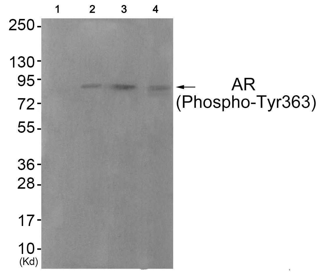
Western blot analysis of extracts from HuvEc cells (Lane 2), COS7 cells (Lane 3) and JK cells (Lane 4), using Androgen Receptor (Phospho-Tyr363) Antibody. The lane on the left is treated with antigen-specific peptide.| Product Name | Androgen Receptor (phospho Tyr363) Antibody |
|---|---|
| Antibody Type | Primary Antibodies |
| Antigen Alias | ANDR, Androgen receptor, Dihydrotestosterone receptor, NR3C4 |
| Product description | The androgen receptor gene is more than 90 kb long and codes for a protein that has 3 major functional domains: the N-terminal domain, DNA-binding domain, and androgen-binding domain. The protein functions as a steroid-hormone activated transcription factor. Upon binding the hormone ligand, the receptor dissociates from accessory proteins, translocates into the nucleus, dimerizes, and then stimulates transcription of androgen responsive genes. This gene contains 2 polymorphic trinucleotide repeat segments that encode polyglutamine and polyglycine tracts in the N-terminal transactivation domain of its protein. Expansion of the polyglutamine tract causes spinal bulbar muscular atrophy (Kennedy disease). Mutations in this gene are also associated with complete androgen insensitivity (CAIS). Two alternatively spliced variants encoding distinct isoforms have been described.1) Lubahn D.B., Mol. Endocrinol. 2:1265-1275(1988). |
| Immunogen | Androgen Receptor (phospho Tyr363) antibody was raised against a peptide sequence around phosphorylation site of tyrosine 363 (D-Y-Y (p)-N-F) derived from Human Androgen Receptor. |
| Modification | p-Tyr363 |
| Clonality | Polyclonal |
|---|---|
| Isotype | IgG |
| Host Species | Rabbit |
| Tested Applications | WB |
| Western Blot: 1:500~1:1000: |
|
| Species Reactivity | HumanMouse |
| Concentration | 1mg/ml |
| Purification | Affinity purified |
| Gene Symbol | AR |
|---|---|
| Alternative Names | ANDR Androgen receptor Dihydrotestosterone receptor NR3C4 |
| Molecular Weight(MW) | 85 kDa |
| Tissue Specificity | The antibody detects endogenous levels of Androgen Receptor only when phosphorylated at tyrosine 363. |
| Entrez Gene | 367 |
|---|---|
| Protein Accession | NP_000035.2 |
Application
Western blot analysis of extracts from HuvEc cells (Lane 2), COS7 cells (Lane 3) and JK cells (Lane 4), using Androgen Receptor (Phospho-Tyr363) Antibody. The lane on the left is treated with antigen-specific peptide.| Application Notes | Western Blot: 1:500~1:1000: |
|---|
| Form | Liquid |
|---|---|
| Storage Instructions | Store antibody at -20˚C for up to one year. |
| Storage Buffer | Antibody supplied in phosphate buffered saline (without Mg2+ and Ca2+), pH 7.4, 150mM NaCl, 0.02% sodium azide and 50% glycerol. |


