Application
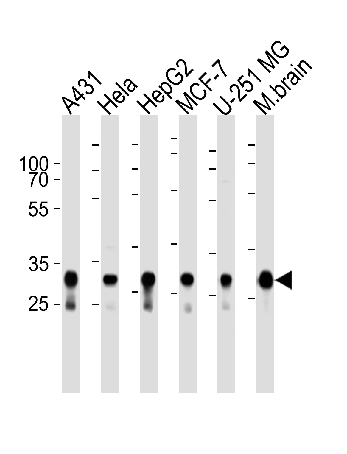
Western blot analysis of lysates from A431, Hela, HepG2, MCF-7, U-251 MG cell lines and mouse brain tissue lysate (from left to right), using CYC1 Antibody at 1:1000 at each lane.Application
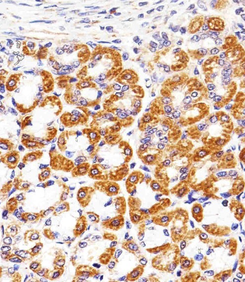
Immunohistochemical analysis of paraffin-embedded R. stomach section using CYC1 Antibody . Antibody was diluted at 1:25 dilution. A peroxidase-conjugated goat anti-rabbit IgG at 1:400 dilution was used as the secondary antibody, followed by DAB staining.Application
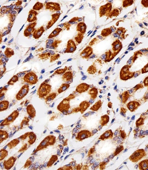
Immunohistochemical analysis of paraffin-embedded H. stomach section using CYC1 Antibody . Antibody was diluted at 1:25 dilution. A peroxidase-conjugated goat anti-rabbit IgG at 1:400 dilution was used as the secondary antibody, followed by DAB staining.Application
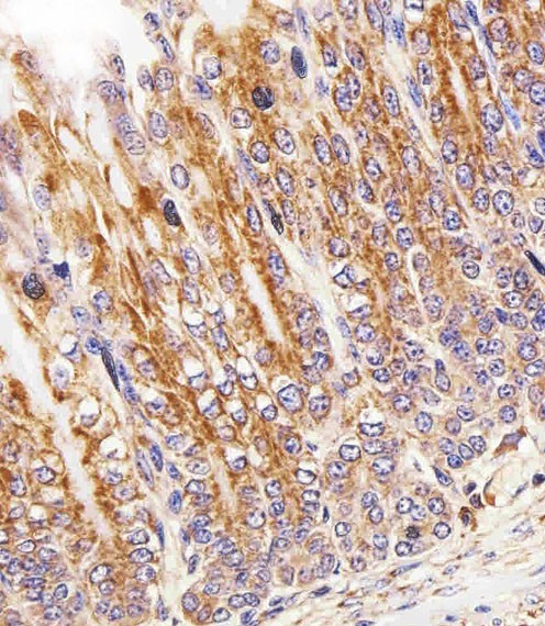
Immunohistochemical analysis of paraffin-embedded M. stomach section using CYC1 Antibody . Antibody was diluted at 1:25 dilution. A peroxidase-conjugated goat anti-rabbit IgG at 1:400 dilution was used as the secondary antibody, followed by DAB staining.Application
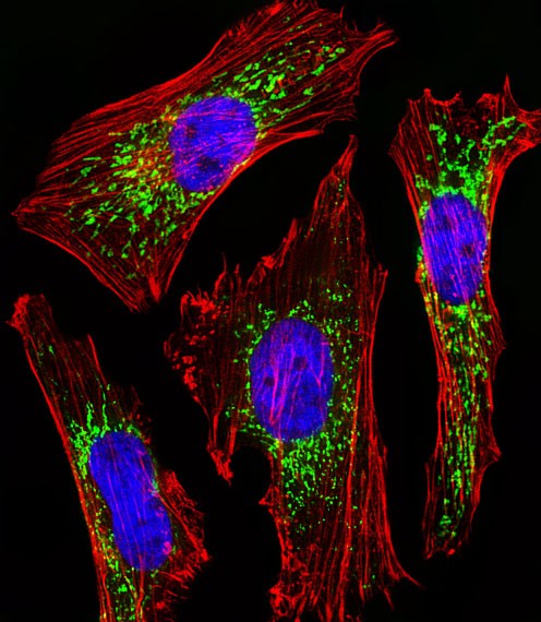
Fluorescent image of Hela cells stained with CYC1 Antibody . Antibody was diluted at 1:25 dilution. An Alexa Fluor 488-conjugated goat anti-rabbit lgG at 1:400 dilution was used as the secondary antibody (green). DAPI was used to stain the cell nuclear (blue). Cytoplasmic actin was counterstained with Alexa Fluor 555 conjugated with Phalloidin (red).| Product Name | CYC1 Antibody |
|---|---|
| Antibody Type | Primary Antibodies |
| Product description | This is the heme-containing component of the cytochrome b-c1 complex, which accepts electrons from Rieske protein and transfers electrons to cytochrome c in the mitochondrial respiratory chain.1) Nishikimi M.,et al.Nucleic Acids Res. 16:3577-3577(1988). |
| Immunogen | This CYC1 antibody is generated from a rabbit immunized with a KLH conjugated synthetic peptide between 142-176 amino acids from the Central region of human CYC1. |
| Clonality | Polyclonal |
|---|---|
| Isotype | Ig |
| Host Species | Rabbit |
| Tested Applications | IFIHC-PWB |
| For IF starting dilution is: 1:25 For IHC-P starting dilution is: 1:25 For WB starting dilution is: 1:1000 | |
| Species Reactivity | HumanMouse |
| Concentration | 1mg/ml |
| Purification | Affinity purified |
| Gene Symbol | CYC1 |
|---|---|
| Alternative Names | Cytochrome c1 heme protein mitochondrial Complex III subunit 4 Complex III subunit IV Cytochrome b-c1 complex subunit 4 Ubiquinol-cytochrome-c reductase complex cytochrome c1 subunit Cytochrome c-1 CYC1 |
| Molecular Weight(MW) | 35 kDa |
Application
Western blot analysis of lysates from A431, Hela, HepG2, MCF-7, U-251 MG cell lines and mouse brain tissue lysate (from left to right), using CYC1 Antibody at 1:1000 at each lane.
Application
Immunohistochemical analysis of paraffin-embedded R. stomach section using CYC1 Antibody . Antibody was diluted at 1:25 dilution. A peroxidase-conjugated goat anti-rabbit IgG at 1:400 dilution was used as the secondary antibody, followed by DAB staining.
Application
Immunohistochemical analysis of paraffin-embedded H. stomach section using CYC1 Antibody . Antibody was diluted at 1:25 dilution. A peroxidase-conjugated goat anti-rabbit IgG at 1:400 dilution was used as the secondary antibody, followed by DAB staining.
Application
Immunohistochemical analysis of paraffin-embedded M. stomach section using CYC1 Antibody . Antibody was diluted at 1:25 dilution. A peroxidase-conjugated goat anti-rabbit IgG at 1:400 dilution was used as the secondary antibody, followed by DAB staining.
Application
Fluorescent image of Hela cells stained with CYC1 Antibody . Antibody was diluted at 1:25 dilution. An Alexa Fluor 488-conjugated goat anti-rabbit lgG at 1:400 dilution was used as the secondary antibody (green). DAPI was used to stain the cell nuclear (blue). Cytoplasmic actin was counterstained with Alexa Fluor 555 conjugated with Phalloidin (red).| Application Notes | For IF starting dilution is: 1:25 For IHC-P starting dilution is: 1:25 For WB starting dilution is: 1:1000 |
|---|
| Form | Liquid |
|---|---|
| Storage Instructions | Store at 4˚C for three months and -20˚C, stable for up to one year. As with all antibodies care should be taken to avoid repeated freeze thaw cycles. Antibodies should not be exposed to prolonged high temperatures. |
| Storage Buffer | Supplied in PBS with 0.09% (W/V) sodium azide. |


