Application
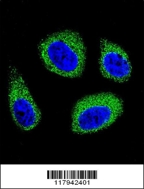
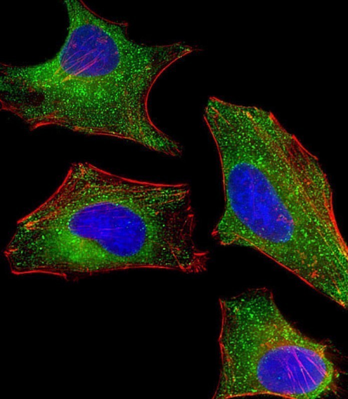
Confocal immunofluorescent analysis of DHCR7 Antibody with MCF-7 cell followed by Alexa Fluor 488-conjugated goat anti-rabbit lgG (green).DAPI was used to stain the cell nuclear (blue).Application
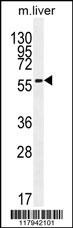
Western blot analysis in mouse liver tissue lysates (35ug/lane).Application
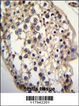
DHCR7 Antibody immunohistochemistry analysis in formalin fixed and paraffin embedded human testis tissue followed by peroxidase conjugation of the secondary antibody and DAB staining.Application
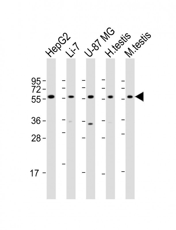
Western Blot at 1:2000 dilution Lane 1: HepG2 whole cell lysate Lane 2: Li-7 whole cell lysate Lane 3: U-87 MG whole cell lysate Lane 4: human testis lysate Lane 5: mouse testis lysate Lysates/proteins at 20 ug per lane.Application
Immunofluorescent analysis of 4% paraformaldehyde-fixed, 0. 1% Triton X-100 permeabilized HeLa (human cervical epithelial adenocarcinoma cell line) cells labeling Pdx1 with antibody at 1/25 dilution, followed by Dylight 488-conjugated goat anti-rabbit IgG (NK179883) secondary antibody at 1/200 dilution (green). Immunofluorescence image showing cytoplasm staining on HeLa cell line. Cytoplasmic acti| Product Name | DHCR7 Antibody |
|---|---|
| Antibody Type | Primary Antibodies |
| Antigen Alias | 7-dehydrocholesterol reductase, 7-DHC reductase, Putative sterol reductase SR-2, Sterol Delta(7)-reductase, DHCR7, D7SR |
| Product description | This gene encodes an enzyme that removes the C(7-8) double bond in the B ring of sterols and catalyzes the conversion of 7-dehydrocholesterol to cholesterol. This gene is ubiquitously expressed and its transmembrane protein localizes to the endoplasmic reticulum membrane and nuclear outer membrane. Mutations in this gene cause Smith-Lemli-Opitz syndrome (SLOS); a syndrome that is metabolically characterized by reduced serum cholesterol levels and elevated serum 7-dehydrocholesterol levels and phenotypically characterized by mental retardation, facial dysmorphism, syndactyly of second and third toes, and holoprosencephaly in severe cases to minimal physical abnormalities and near-normal intelligence in mild cases. Alternative splicing results in multiple transcript variants that encode the same protein.1) Bailey, S.D., et al. Diabetes Care 33(10):2250-2253(2010) |
| Immunogen | This DHCR7 antibody is generated from rabbits immunized with a KLH conjugated synthetic peptide between 437-463 amino acids from the C-terminal region of human DHCR7. |
| Clonality | Polyclonal |
|---|---|
| Isotype | Ig |
| Host Species | Rabbit |
| Tested Applications | IFIHC-PWB |
| For IF starting dilution is: 1:25 For WB starting dilution is: 1:1000 For IHC-P starting dilution is: 1:50~100: |
|
| Species Reactivity | HumanMouse |
| Concentration | 1mg/ml |
| Purification | Affinity purified |
| Gene Symbol | DHCR7 |
|---|---|
| Alternative Names | 7-dehydrocholesterol reductase 7-DHC reductase Putative sterol reductase SR-2 Sterol Delta(7)-reductase DHCR7 D7SR |
| Molecular Weight(MW) | 54 kDa |
Application
Confocal immunofluorescent analysis of DHCR7 Antibody with MCF-7 cell followed by Alexa Fluor 488-conjugated goat anti-rabbit lgG (green).DAPI was used to stain the cell nuclear (blue).
Application
Western blot analysis in mouse liver tissue lysates (35ug/lane).
Application
DHCR7 Antibody immunohistochemistry analysis in formalin fixed and paraffin embedded human testis tissue followed by peroxidase conjugation of the secondary antibody and DAB staining.
Application
Western Blot at 1:2000 dilution Lane 1: HepG2 whole cell lysate Lane 2: Li-7 whole cell lysate Lane 3: U-87 MG whole cell lysate Lane 4: human testis lysate Lane 5: mouse testis lysate Lysates/proteins at 20 ug per lane.
Application
Immunofluorescent analysis of 4% paraformaldehyde-fixed, 0. 1% Triton X-100 permeabilized HeLa (human cervical epithelial adenocarcinoma cell line) cells labeling Pdx1 with antibody at 1/25 dilution, followed by Dylight 488-conjugated goat anti-rabbit IgG (NK179883) secondary antibody at 1/200 dilution (green). Immunofluorescence image showing cytoplasm staining on HeLa cell line. Cytoplasmic acti| Application Notes | For IF starting dilution is: 1:25 For WB starting dilution is: 1:1000 For IHC-P starting dilution is: 1:50~100: |
|---|
| Form | Liquid |
|---|---|
| Storage Instructions | Store at 4˚C for three months and -20˚C, stable for up to one year. As with all antibodies care should be taken to avoid repeated freeze thaw cycles. Antibodies should not be exposed to prolonged high temperatures. |
| Storage Buffer | Supplied in PBS with 0.09% (W/V) sodium azide. |


