Application
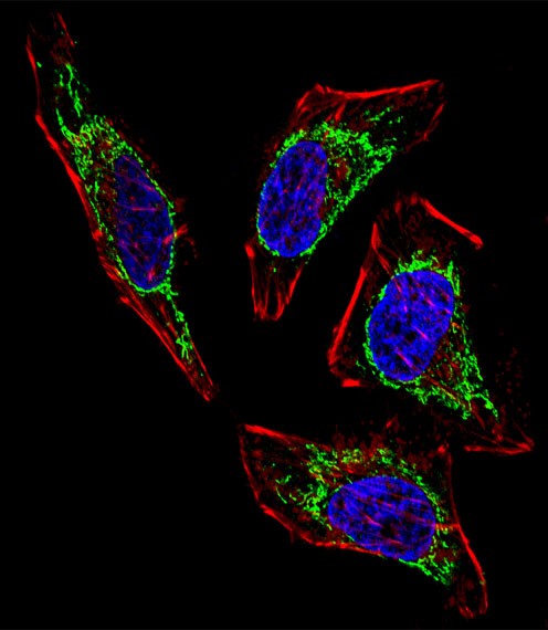
Fluorescent confocal image of Hela cell stained with HSPD1 Antibody . Hela cells were fixed with 4% PFA (20 min), permeabilized with Triton X-100 (0.1%, 10 min), then incubated with HSPD1 primary antibody (1:25). For secondary antibody, Alexa Fluor 488 conjugated donkey anti-rabbit antibody (green) was used (1:400).Cytoplasmic actin was counterstained with Alexa Fluor 555 (red) conjugated PhalloidApplication
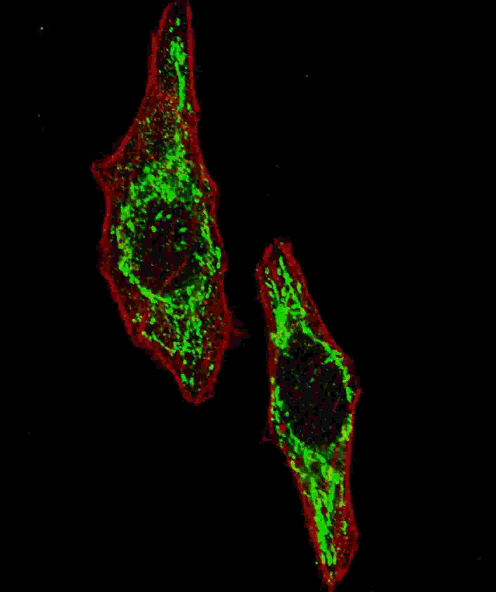
Fluorescent image of U251 cells stained with HSPD1 antibody. U251 cells were fixed with 4% PFA (20 min), permeabilized with Triton X-100 (0.2%, 30 min). Cells were then incubated with HSPD1 primary antibody (1:100, 2 h at room temperature). For secondary antibody, Alexa Fluor 488 conjugated donkey anti-rabbit antibody (green) was used (1:1000, 1h). Cytoplasmic actin was counterstained with Alexa FApplication
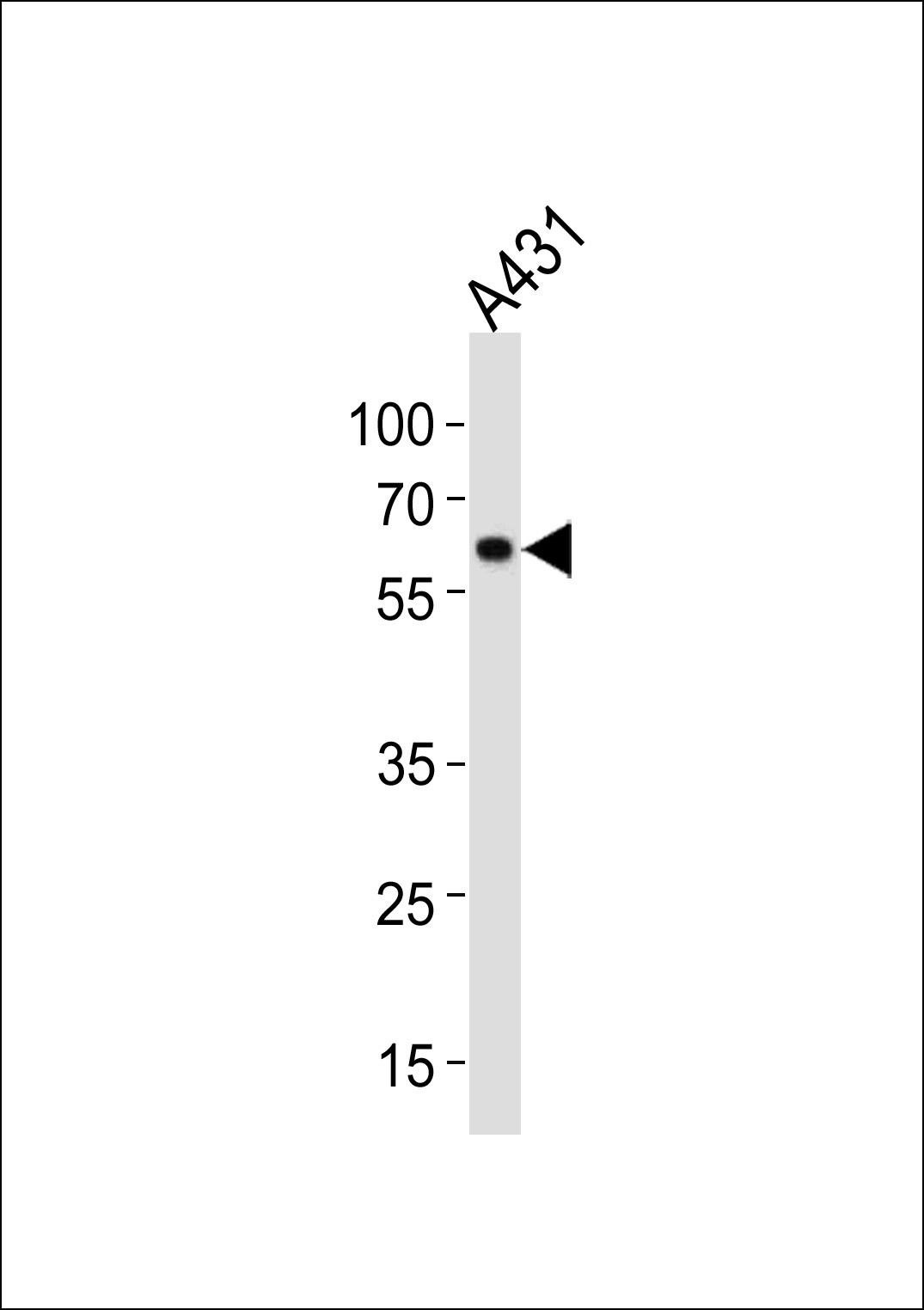
Western blot analysis in A431 cell line lysates (35ug/lane).| Product Name | HSPD1 Antibody |
|---|---|
| Antibody Type | Primary Antibodies |
| Antigen Alias | 60 kDa heat shock protein, mitochondrial, 60 kDa chaperonin, Chaperonin 60, CPN60, Heat shock protein 60, HSP-60, Hsp60, HuCHA60, Mitochondrial matrix protein P1, P60 lymphocyte protein, HSPD1, HSP60 |
| Product description | HSPD1 is a member of the chaperonin family. This protein may function as a signaling molecule in the innate immune system. The protein is essential for the folding and assembly of newly imported proteins in the mitochondria. The protein is adjacent to a related family member and the region between the 2 genes functions as a bidirectional promoter.1) References for protein: |
| Immunogen | This HSPD1 antibody is generated from rabbits immunized with a KLH conjugated synthetic peptide between 187-215 amino acids from the Central region of human HSPD1. |
| Clonality | Polyclonal |
|---|---|
| Isotype | Ig |
| Host Species | Rabbit |
| Tested Applications | IFWB |
| For WB starting dilution is: 1:1000 For IF starting dilution is: 1:100: |
|
| Species Reactivity | Human |
| Concentration | 1mg/ml |
| Gene Symbol | HSPD1 |
|---|---|
| Alternative Names | 60 kDa heat shock protein mitochondrial 60 kDa chaperonin Chaperonin 60 CPN60 Heat shock protein 60 HSP-60 Hsp60 HuCHA60 Mitochondrial matrix protein P1 P60 lymphocyte protein HSPD1 HSP60 |
| Molecular Weight(MW) | 61 kDa |
| Sequence Similarities | Predicted species reactivity based on immunogen sequence: Bovine |
Application
Fluorescent confocal image of Hela cell stained with HSPD1 Antibody . Hela cells were fixed with 4% PFA (20 min), permeabilized with Triton X-100 (0.1%, 10 min), then incubated with HSPD1 primary antibody (1:25). For secondary antibody, Alexa Fluor 488 conjugated donkey anti-rabbit antibody (green) was used (1:400).Cytoplasmic actin was counterstained with Alexa Fluor 555 (red) conjugated Phalloid
Application
Fluorescent image of U251 cells stained with HSPD1 antibody. U251 cells were fixed with 4% PFA (20 min), permeabilized with Triton X-100 (0.2%, 30 min). Cells were then incubated with HSPD1 primary antibody (1:100, 2 h at room temperature). For secondary antibody, Alexa Fluor 488 conjugated donkey anti-rabbit antibody (green) was used (1:1000, 1h). Cytoplasmic actin was counterstained with Alexa F
Application
Western blot analysis in A431 cell line lysates (35ug/lane).| Application Notes | For WB starting dilution is: 1:1000 For IF starting dilution is: 1:100: |
|---|
| Form | Liquid |
|---|---|
| Storage Instructions | Store at 4˚C for three months and -20˚C, stable for up to one year. As with all antibodies care should be taken to avoid repeated freeze thaw cycles. Antibodies should not be exposed to prolonged high temperatures. |
| Storage Buffer | Supplied in PBS with 0.09% (W/V) sodium azide. |


