Application
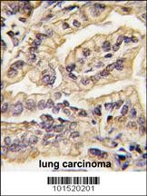
Formalin-fixed and paraffin-embedded human lung carcinoma tissue reacted with CSF1R Antibody , which was peroxidase-conjugated to the secondary antibody, followed by DAB staining.Application
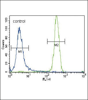
Flow cytometric analysis of NCI-H460 cells (right histogram) compared to a negative control cell (left histogram).FITC-conjugated goat-anti-rabbit secondary antibodies were used for the analysis.Application
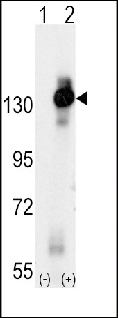
Western blot analysis of CSF1R using rabbit polyclonal CSF1R Antibody using 293 cell lysates (2 ug/lane) either nontransfected (Lane 1) or transiently transfected with the CSF1R gene (Lane 2).Application
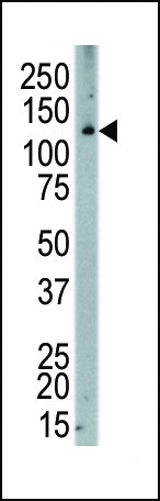
Western blot analysis of anti-CSF1R Pab in human placenta| Product Name | MCSF Receptor Antibody |
|---|---|
| Antibody Type | Primary Antibodies |
| Product description | CSF1R is the receptor for colony stimulating factor 1, a cytokine which controls the production, differentiation, and function of macrophages. This receptor mediates most if not all of the biological effects of this cytokine. Ligand binding activates the receptor kinase through a process of oligomerization and transphosphorylation. This protein is a tyrosine kinase transmembrane receptor and member of the CSF1/PDGF receptor family of tyrosine-protein kinases. Mutations in the gene encoding CSF1R have been associated with a predisposition to myeloid malignancy.1) Follows, G.A., et al., EMBO J. 22(11):2798-2809 (2003). |
| Immunogen | This MCSF Receptor (CSF1R) antibody is generated from rabbits immunized with a KLH conjugated synthetic peptide between 940-971 amino acids from the C-terminal region of human MCSF Receptor (CSF1R). |
| Clonality | Polyclonal |
|---|---|
| Isotype | Ig |
| Host Species | Rabbit |
| Tested Applications | FACSIHC-PWB |
| For WB starting dilution is: 1:1000 For FACS starting dilution is: 1:10~50 For IHC-P starting dilution is: 1:10~50 | |
| Species Reactivity | Human |
| Concentration | 1mg/ml |
| Purification | Unpurified |
| Gene Symbol | CSF1R |
|---|---|
| Alternative Names | Macrophage colony-stimulating factor 1 receptor CSF-1 receptor CSF-1-R CSF-1R M-CSF-R Proto-oncogene c-Fms CD115 CSF1R FMS |
| Molecular Weight(MW) | 108 kDa |
Application
Formalin-fixed and paraffin-embedded human lung carcinoma tissue reacted with CSF1R Antibody , which was peroxidase-conjugated to the secondary antibody, followed by DAB staining.
Application
Flow cytometric analysis of NCI-H460 cells (right histogram) compared to a negative control cell (left histogram).FITC-conjugated goat-anti-rabbit secondary antibodies were used for the analysis.
Application
Western blot analysis of CSF1R using rabbit polyclonal CSF1R Antibody using 293 cell lysates (2 ug/lane) either nontransfected (Lane 1) or transiently transfected with the CSF1R gene (Lane 2).
Application
Western blot analysis of anti-CSF1R Pab in human placenta| Application Notes | For WB starting dilution is: 1:1000 For FACS starting dilution is: 1:10~50 For IHC-P starting dilution is: 1:10~50 |
|---|
| Form | Liquid |
|---|---|
| Storage Instructions | Store at 4˚C for three months and -20˚C, stable for up to one year. As with all antibodies care should be taken to avoid repeated freeze thaw cycles. Antibodies should not be exposed to prolonged high temperatures. |
| Storage Buffer | Supplied in PBS with 0.09% (W/V) sodium azide. |


