Application
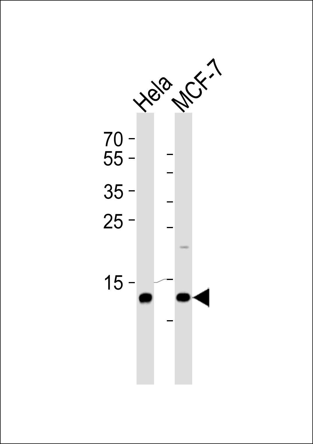
Western blot analysis of lysates from Hela, MCF-7 cell line (from left to right), using RBX1 Antibody at 1:1000 at each lane.Application
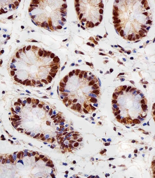
Immunohistochemical analysis of paraffin-embedded H. colon section using RBX1 Antibody . Antibody was diluted at 1:100 dilution. A peroxidase-conjugated goat anti-rabbit IgG at 1:400 dilution was used as the secondary antibody, followed by DAB staining.Application
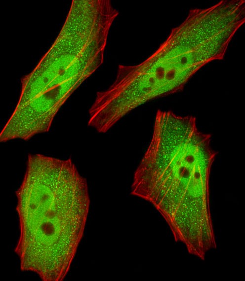
Fluorescent image of HeLa cells stained with RBX1 Antibody . Antibody was diluted at 1:25 dilution. An Alexa Fluor 488-conjugated goat anti-rabbit lgG at 1:400 dilution was used as the secondary antibody (green). Cytoplasmic actin was counterstained with Alexa Fluor 555 conjugated with Phalloidin (red).Application
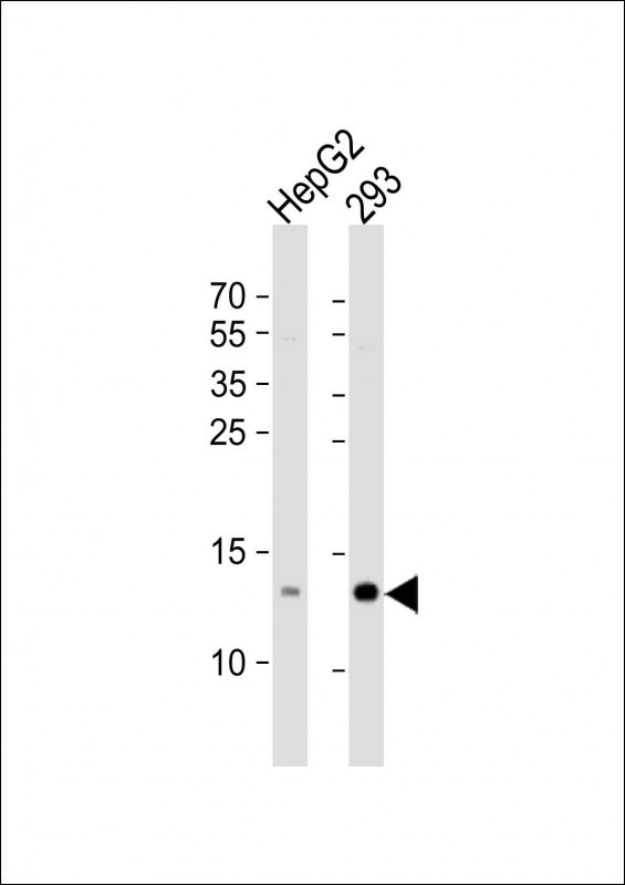
Western Blot at 1:1000 dilution Lane 1: HepG2 whole cell lysate Lane 2: 293 whole cell lysate Lysates/proteins at 20 ug per lane.Application
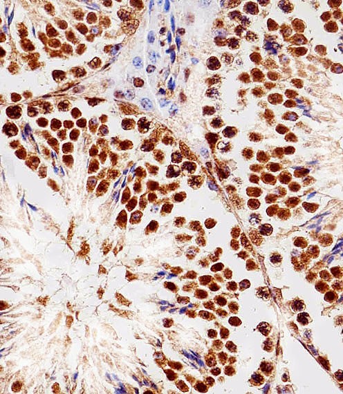
Immunohistochemical analysis of paraffin-embedded M. testis section using RBX1 Antibody . Antibody was diluted at 1:100 dilution. A peroxidase-conjugated goat anti-rabbit IgG at 1:400 dilution was used as the secondary antibody, followed by DAB staining.| Product Name | RBX1 Antibody |
|---|---|
| Antibody Type | Primary Antibodies |
| Antigen Alias | E3 ubiquitin-protein ligase RBX1, 632-, Protein ZYP, RING finger protein 75, RING-box protein 1, Rbx1, Regulator of cullins 1, E3 ubiquitin-protein ligase RBX1, N-terminally processed, RBX1, RNF75, ROC1 |
| Product description | E3 ubiquitin ligase component of multiple cullin-RING- based E3 ubiquitin-protein ligase complexes which mediate the ubiquitination and subsequent proteasomal degradation of target proteins, including proteins involved in cell cycle progression, signal transduction, transcription and transcription-coupled nucleotide excision repair. The functional specificity of the E3 ubiquitin-protein ligase complexes depends on the variable substrate recognition components. As a component of the CSA complex promotes the ubiquitination of ERCC6 resulting in proteasomal degradation. Through the RING-type zinc finger, seems to recruit the E2 ubiquitination enzyme, like CDC34, to the complex and brings it into close proximity to the substrate. Probably also stimulates CDC34 autoubiquitination. May be required for histone H3 and histone H4 ubiquitination in response to ultraviolet and for subsequent DNA repair. Promotes the neddylation of CUL1, CUL2, CUL4 and CUL4 via its interaction with UBE2M. Involved in the ubiquitination of KEAP1, ENC1 and KLHL41. In concert with ATF2 and CUL3, promotes degradation of KAT5 thereby attenuating its ability to acetylate and activate ATM.1) Ohta T.,et al.Mol. Cell 3:535-541(1999). |
| Immunogen | This RBX1 antibody is generated from a rabbit immunized with a KLH conjugated synthetic peptide between 74-108 amino acids from the C-terminal region of human RBX1. |
| Clonality | Polyclonal |
|---|---|
| Isotype | Ig |
| Host Species | Rabbit |
| Tested Applications | IFIHC-PWB |
| For WB starting dilution is: 1:1000 For IHC-P starting dilution is: 1:25 For IF starting dilution is: 1:25: |
|
| Species Reactivity | Human |
| Concentration | 1mg/ml |
| Purification | Affinity purified |
| Gene Symbol | RBX1 |
|---|---|
| Alternative Names | E3 ubiquitin-protein ligase RBX1 632- Protein ZYP RING finger protein 75 RING-box protein 1 Rbx1 Regulator of cullins 1 E3 ubiquitin-protein ligase RBX1 N-terminally processed RBX1 RNF75 ROC1 |
| Molecular Weight(MW) | 12 kDa |
| Sequence Similarities | Predicted species reactivity based on immunogen sequence: C. Elegans |
Application
Western blot analysis of lysates from Hela, MCF-7 cell line (from left to right), using RBX1 Antibody at 1:1000 at each lane.
Application
Immunohistochemical analysis of paraffin-embedded H. colon section using RBX1 Antibody . Antibody was diluted at 1:100 dilution. A peroxidase-conjugated goat anti-rabbit IgG at 1:400 dilution was used as the secondary antibody, followed by DAB staining.
Application
Fluorescent image of HeLa cells stained with RBX1 Antibody . Antibody was diluted at 1:25 dilution. An Alexa Fluor 488-conjugated goat anti-rabbit lgG at 1:400 dilution was used as the secondary antibody (green). Cytoplasmic actin was counterstained with Alexa Fluor 555 conjugated with Phalloidin (red).
Application
Western Blot at 1:1000 dilution Lane 1: HepG2 whole cell lysate Lane 2: 293 whole cell lysate Lysates/proteins at 20 ug per lane.
Application
Immunohistochemical analysis of paraffin-embedded M. testis section using RBX1 Antibody . Antibody was diluted at 1:100 dilution. A peroxidase-conjugated goat anti-rabbit IgG at 1:400 dilution was used as the secondary antibody, followed by DAB staining.| Application Notes | For WB starting dilution is: 1:1000 For IHC-P starting dilution is: 1:25 For IF starting dilution is: 1:25: |
|---|
| Form | Liquid |
|---|---|
| Storage Instructions | Store at 4˚C for three months and -20˚C, stable for up to one year. As with all antibodies care should be taken to avoid repeated freeze thaw cycles. Antibodies should not be exposed to prolonged high temperatures. |
| Storage Buffer | Supplied in PBS with 0.09% (W/V) sodium azide. |


