Application
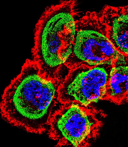
Fluorescent confocal image of A431 cell stained with RCAN1 Antibody (N-term).A431 cells were fixed with 4% PFA (20 min), permeabilized with Triton X-100 (0.1%, 10 min), then incubated with RCAN1 primary antibody (1:25). For secondary antibody, Alexa Fluor 488 conjugated donkey anti-rabbit antibody (green) was used (1:400).Cytoplasmic actin was counterstained with Alexa Fluor 555 (red) conjugated PApplication
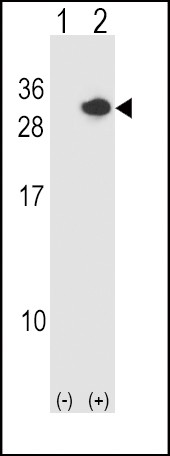
Western blot analysis of RCAN1 using rabbit polyclonal RCAN1 Antibody using 293 cell lysates (2 ug/lane) either nontransfected (Lane 1) or transiently transfected (Lane 2) with the RCAN1 gene.Application
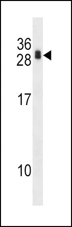
Western blot analysis in 293 cell line lysates (35ug/lane).| Product Name | RCAN1 Antibody |
|---|---|
| Antibody Type | Primary Antibodies |
| Antigen Alias | Calcipressin-1, Adapt78, Down syndrome critical region protein 1, Myocyte-enriched calcineurin-interacting protein 1, MCIP1, Regulator of calcineurin 1, RCAN1, ADAPT78, CSP1, DSC1, DSCR1 |
| Product description | The protein encoded by this gene interacts with calcineurin A and inhibits calcineurin-dependent signaling pathways, possibly affecting central nervous system development. This gene is located in the minimal candidate region for the Down syndrome phenotype, and is overexpressed in the brain of Down syndrome fetuses. Chronic overexpression of this gene may lead to neurofibrillary tangles such as those associated with Alzheimer disease. Three transcript variants encoding three different isoforms have been found for this gene.1) Rose, J.E., et al. Mol. Med. 16 (7-8), 247-253 (2010) : |
| Immunogen | This RCAN1 antibody is generated from rabbits immunized with a KLH conjugated synthetic peptide between 41-68 amino acids from the N-terminal region of human RCAN1. |
| Clonality | Polyclonal |
|---|---|
| Isotype | Ig |
| Host Species | Rabbit |
| Tested Applications | IFWB |
| For WB starting dilution is: 1:1000 For IF starting dilution is: 1:10~50: |
|
| Species Reactivity | Human |
| Concentration | 1mg/ml |
| Purification | Affinity purified |
| Gene Symbol | RCAN1 |
|---|---|
| Alternative Names | Calcipressin-1 Adapt78 Down syndrome critical region protein 1 Myocyte-enriched calcineurin-interacting protein 1 MCIP1 Regulator of calcineurin 1 RCAN1 ADAPT78 CSP1 DSC1 DSCR1 |
| Molecular Weight(MW) | 28 kDa |
| Sequence Similarities | Predicted species reactivity based on immunogen sequence: Mouse |
Application
Fluorescent confocal image of A431 cell stained with RCAN1 Antibody (N-term).A431 cells were fixed with 4% PFA (20 min), permeabilized with Triton X-100 (0.1%, 10 min), then incubated with RCAN1 primary antibody (1:25). For secondary antibody, Alexa Fluor 488 conjugated donkey anti-rabbit antibody (green) was used (1:400).Cytoplasmic actin was counterstained with Alexa Fluor 555 (red) conjugated P
Application
Western blot analysis of RCAN1 using rabbit polyclonal RCAN1 Antibody using 293 cell lysates (2 ug/lane) either nontransfected (Lane 1) or transiently transfected (Lane 2) with the RCAN1 gene.
Application
Western blot analysis in 293 cell line lysates (35ug/lane).| Application Notes | For WB starting dilution is: 1:1000 For IF starting dilution is: 1:10~50: |
|---|
| Form | Liquid |
|---|---|
| Storage Instructions | Store at 4˚C for three months and -20˚C, stable for up to one year. As with all antibodies care should be taken to avoid repeated freeze thaw cycles. Antibodies should not be exposed to prolonged high temperatures. |
| Storage Buffer | Supplied in PBS with 0.09% (W/V) sodium azide. |


