Application
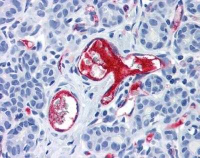
Immunohistochemistry staining of VWF in breast, vessels tissue using VWF monoclonal Antibody.WB
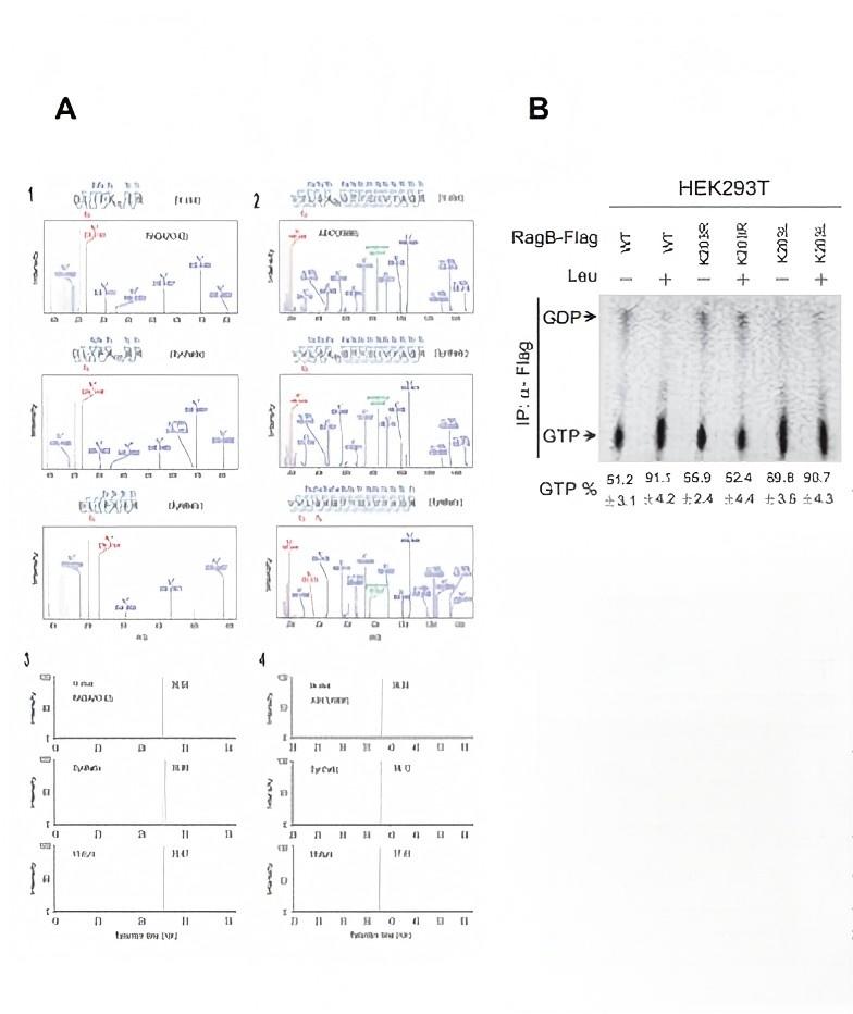
(A, B) Representative image of CD31 (A) and vWF (B) staining (red) in the lesion epicenter and colocalization with the transplanted GFP-MSCs (green).| Product Name | VWF Antibody |
|---|---|
| Antibody Type | Primary Antibodies |
| Immunogen | VWF monoclonal antibody was raised against human Factor VIII (VWF) (Human). |
| Clonality | Monoclonal |
|---|---|
| Isotype | IgG1 |
| Host Species | Mouse |
| Tested Applications | ELISAIFIHCIPRIAWB |
| VWF antibody can be used in immunohistochemistry starting at 20 μg/mL.: |
|
| Species Reactivity | Human |
| Concentration | 1mg/ml |
| Purification | Affinity purified |
| Gene Symbol | VWF |
|---|---|
| Alternative Names | VWF Coagulation factor VIII VWF F8VWF VWD Von Willebrand factor |
| Tissue Specificity | Reacts strongly with human von Willebrand Factor or Factor VIII Related Antigen in both plasma and tissues. |
Application
Immunohistochemistry staining of VWF in breast, vessels tissue using VWF monoclonal Antibody.
WB
(A, B) Representative image of CD31 (A) and vWF (B) staining (red) in the lesion epicenter and colocalization with the transplanted GFP-MSCs (green).| Application Notes | VWF antibody can be used in immunohistochemistry starting at 20 μg/mL.: |
|---|
| Form | Liquid |
|---|---|
| Storage Instructions | VWF antibody can be stored short term 4 ˚C. For long term storage aliquot and store at -20 ˚C. As with all antibodies avoid freeze/thaw cycles. |
| Storage Buffer | PBS, pH 7.4, 0.1% sodium azide, 0.2% BSA, 50% glycerol |


