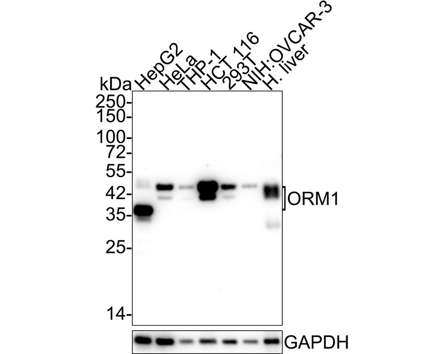


WB
Western blot analysis of Alpha 1 Acid Glycoprotein on different lysates with Mouse anti-Alpha 1 Acid Glycoprotein antibody at 1/1,000 dilution. Lane 1: HepG2 cell lysate (20 µg/Lane), Lane 2: HeLa cell lysate (20 µg/Lane), Lane 3: THP-1 cell lysate (20 µg/Lane), Lane 4: HCT 116 cell lysate (20 µg/Lane), Lane 5: 293T cell lysate (20 µg/Lane), Lane 6: NIH:OVCAR-3 cell lysate (20 µg/Lane), Lane 7: Human liver tissue lysate (40 µg/Lane), Exposure time: 1 minute 59 seconds; 4-20% SDS-PAGE gel. Proteins were transferred to a PVDF membrane and blocked with 5% NFDM/TBST for 1 hour at room temperature. The primary antibody at 1/1,000 dilution was used in 5% NFDM/TBST at 4℃ overnight. Goat Anti-Mouse IgG - HRP Secondary Antibody at 1/50,000 dilution was used for 1 hour at room temperature.IHC
Immunohistochemical analysis of paraffin-embedded rat liver tissue using anti- Alpha-1-acid glycoprotein antibody. Counter stained with hematoxylin.IHC
Immunohistochemical analysis of paraffin-embedded human lung cancer tissue using anti-Alpha-1-acid glycoprotein antibody. Counter stained with hematoxylin.IHC
Immunohistochemical analysis of paraffin-embedded human esophagus tissue using anti-Alpha-1-acid glycoprotein antibody. Counter stained with hematoxylin.FC
Flow cytometric analysis of HepG2 cells with Alpha-1-acid glycoprotein antibody at 1/100 dilution (purple) compared with an unlabelled control (cells without incubation with primary antibody; yellow). Alexa Fluor 488-conjugated goat anti-rabbit IgG was used as the secondary antibody.| Product Name | Alpha 1 Acid Glycoprotein Mouse Monoclonal Antibody |
|---|---|
| Antibody Type | Primary Antibodies |
| Product description | Functions as transport protein in the blood stream. Binds various ligands in the interior of its beta-barrel domain. Also binds synthetic drugs and influences their distribution and availability in the body. Appears to function in modulating the activity of the immune system during the acute-phase reaction. |
| Immunogen | Recombinant protein within Human ORM1 aa 1-201 / 201. |
| Clonality | Monoclonal |
|---|---|
| Isotype | IgG1 |
| Host Species | Mouse |
| WB:1:1000 IHC:1:50-1:100 FC:1:50-1:100 |
|
| Concentration | 2mg/ml |
| Purification | Protein G |
| Alternative Names | A1AG1_HUMAN antibody
AGP 1 antibody
AGP A antibody
AGP antibody
AGP1 antibody
Alpha 1 acid glycoprotein antibody
Alpha-1-acid glycoprotein 1 antibody
alpha-1-AGP antibody
Epididymis secretory sperm binding protein Li 153w antibody
glycoprotein alpha-1-acid of serum antibody HEL S 153w antibody OMD 1 antibody ORM antibody ORM1 antibody Orosomucoid 1 antibody Orosomucoid-1 antibody |
|---|---|
| Molecular Weight(MW) | 24 kDa (Observed band size: 37/45 kDa) |
| Cellular Localization | Secreted. |
WB
Western blot analysis of Alpha 1 Acid Glycoprotein on different lysates with Mouse anti-Alpha 1 Acid Glycoprotein antibody at 1/1,000 dilution. Lane 1: HepG2 cell lysate (20 µg/Lane), Lane 2: HeLa cell lysate (20 µg/Lane), Lane 3: THP-1 cell lysate (20 µg/Lane), Lane 4: HCT 116 cell lysate (20 µg/Lane), Lane 5: 293T cell lysate (20 µg/Lane), Lane 6: NIH:OVCAR-3 cell lysate (20 µg/Lane), Lane 7: Human liver tissue lysate (40 µg/Lane), Exposure time: 1 minute 59 seconds; 4-20% SDS-PAGE gel. Proteins were transferred to a PVDF membrane and blocked with 5% NFDM/TBST for 1 hour at room temperature. The primary antibody at 1/1,000 dilution was used in 5% NFDM/TBST at 4℃ overnight. Goat Anti-Mouse IgG - HRP Secondary Antibody at 1/50,000 dilution was used for 1 hour at room temperature.
IHC
Immunohistochemical analysis of paraffin-embedded rat liver tissue using anti- Alpha-1-acid glycoprotein antibody. Counter stained with hematoxylin.
IHC
Immunohistochemical analysis of paraffin-embedded human lung cancer tissue using anti-Alpha-1-acid glycoprotein antibody. Counter stained with hematoxylin.
IHC
Immunohistochemical analysis of paraffin-embedded human esophagus tissue using anti-Alpha-1-acid glycoprotein antibody. Counter stained with hematoxylin.
FC
Flow cytometric analysis of HepG2 cells with Alpha-1-acid glycoprotein antibody at 1/100 dilution (purple) compared with an unlabelled control (cells without incubation with primary antibody; yellow). Alexa Fluor 488-conjugated goat anti-rabbit IgG was used as the secondary antibody.| Application Notes | WB:1:1000 IHC:1:50-1:100 FC:1:50-1:100 |
|---|
| Form | Liquid |
|---|---|
| Storage Instructions | Store at +4℃ after thawing. Aliquot store at -20℃. Avoid repeated freeze / thaw cycles. |
| Storage Buffer | 1*TBS (pH7.4), 0.5%BSA, 50%Glycerol. Preservative: 0.05% Sodium Azide. |


