

Application
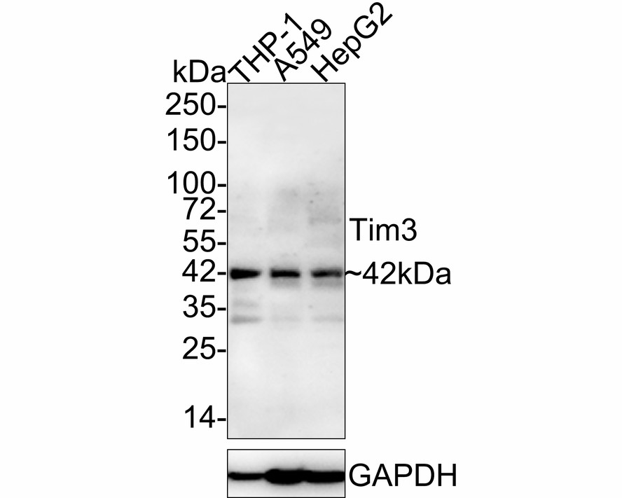
Fig1: Western blot analysis of TIM 3 on different lysates with Mouse anti-TIM 3 antibody at 1/2,000 dilution. Lane 1: THP-1 cell lysate (no heat), Lane 2: A549 cell lysate (no heat), Lane 3: HepG2 cell lysate (no heat), Notice: no heat means the lysate is not boiled. Lysates/proteins at 30 µg/Lane. Exposure time: 5 minutes 20 seconds; 4-20% SDS-PAGE gel. Proteins were transferred to a PVDF membrane and blocked with 5% NFDM/TBST for 1 hour at room temperature. The primary antibody at 1/2,000 dilution was used in 5% NFDM/TBST at room temperature for 2 hours. Goat Anti-Mouse IgG - HRP Secondary Antibody at 1/50,000 dilution was used for 1 hour at room temperature.Application
Fig2: ICC staining Tim3 (green) in Hela cells. The nuclear counter stain is DAPI (blue). Cells were fixed in paraformaldehyde, permeabilised with 0.25% Triton X100/PBS.Application
Fig3: ICC staining Tim3 (green) in 293T cells. The nuclear counter stain is DAPI (blue). Cells were fixed in paraformaldehyde, permeabilised with 0.25% Triton X100/PBS.Application
Fig4: Immunohistochemical analysis of paraffin-embedded human lung cancer tissue using anti-Tim3 antibody. Counter stained with hematoxylin.Application
Fig5: Immunohistochemical analysis of paraffin-embedded human spleen tissue using anti-Tim3 antibody. Counter stained with hematoxylin.Application
Fig6: Immunohistochemical analysis of paraffin-embedded human tonsil tissue using anti-Tim3 antibody. Counter stained with hematoxylin.Application
Fig7: Flow cytometric analysis of Daudi cells with Tim3 antibody at 1/100 dilution (red) compared with an unlabelled control (cells without incubation with primary antibody; black).| Product Name | TIM 3 Mouse Monoclonal Antibody |
|---|---|
| Antibody Type | Primary Antibodies |
| Product description | Cell surface receptor implicated in modulating innate and adaptive immune responses. Generally accepted to have an inhibiting function. Reports on stimulating functions suggest that the activity may be influenced by the cellular context and/or the respective ligand. Regulates macrophage activation. Inhibits T-helper type 1 lymphocyte (Th1)-mediated auto- and alloimmune responses and promotes immunological tolerance. In CD8+ cells attenuates TCR-induced signaling, specifically by blocking NF-kappaB and NFAT promoter activities resulting in the loss of IL-2 secretion. The function may implicate its association with LCK proposed to impair phosphorylation of TCR subunits, and/or LGALS9-dependent recruitment of PTPRC to the immunological synapse. |
| Immunogen | Recombinant protein with Human TIM 3 1-89 / 301. |
| Clonality | Monoclonal |
|---|---|
| Isotype | IgG1 |
| Host Species | Mouse |
| Tested Applications | FCICC/IFIHCWB |
| WB:1:500-1:2000 IHC:1:100-1:200 ICC/IF:1:50-1:200 FC:1:50-1:100 |
|
| Species Reactivity | Human |
| Concentration | 2mg/ml |
| Purification | Protein A |
| Alternative Names | CD366 antibody FLJ14428 antibody HAVcr-2 antibody Havcr2 antibody HAVR2_HUMAN antibody Hepatitis A virus cellular receptor 2 antibody Kidney injury molecule 3 antibody KIM 3 antibody KIM3 antibody T cell immunoglobulin and mucin domain containing 3 antibody T cell immunoglobulin mucin 3 antibody T-cell immunoglobulin and mucin domain-containing protein 3 antibody T-cell immunoglobulin mucin family member 3 antibody T-cell immunoglobulin mucin receptor 3 antibody T-cell membrane protein 3 antibody Tim 3 antibody TIM-3 antibody TIM3 antibody TIMD-3 antibody TIMD3 antibody |
|---|---|
| Molecular Weight(MW) | 33 kDa(Observed band size: 42 kDa) |
| Cellular Localization | Membrane. Cell junction. |
| SwissProt ID | Q8TDQ0 |
|---|
Application
Fig1: Western blot analysis of TIM 3 on different lysates with Mouse anti-TIM 3 antibody at 1/2,000 dilution. Lane 1: THP-1 cell lysate (no heat), Lane 2: A549 cell lysate (no heat), Lane 3: HepG2 cell lysate (no heat), Notice: no heat means the lysate is not boiled. Lysates/proteins at 30 µg/Lane. Exposure time: 5 minutes 20 seconds; 4-20% SDS-PAGE gel. Proteins were transferred to a PVDF membrane and blocked with 5% NFDM/TBST for 1 hour at room temperature. The primary antibody at 1/2,000 dilution was used in 5% NFDM/TBST at room temperature for 2 hours. Goat Anti-Mouse IgG - HRP Secondary Antibody at 1/50,000 dilution was used for 1 hour at room temperature.
Application
Fig2: ICC staining Tim3 (green) in Hela cells. The nuclear counter stain is DAPI (blue). Cells were fixed in paraformaldehyde, permeabilised with 0.25% Triton X100/PBS.
Application
Fig3: ICC staining Tim3 (green) in 293T cells. The nuclear counter stain is DAPI (blue). Cells were fixed in paraformaldehyde, permeabilised with 0.25% Triton X100/PBS.
Application
Fig4: Immunohistochemical analysis of paraffin-embedded human lung cancer tissue using anti-Tim3 antibody. Counter stained with hematoxylin.
Application
Fig5: Immunohistochemical analysis of paraffin-embedded human spleen tissue using anti-Tim3 antibody. Counter stained with hematoxylin.
Application
Fig6: Immunohistochemical analysis of paraffin-embedded human tonsil tissue using anti-Tim3 antibody. Counter stained with hematoxylin.
Application
Fig7: Flow cytometric analysis of Daudi cells with Tim3 antibody at 1/100 dilution (red) compared with an unlabelled control (cells without incubation with primary antibody; black).| Application Notes | WB:1:500-1:2000 IHC:1:100-1:200 ICC/IF:1:50-1:200 FC:1:50-1:100 |
|---|
| Form | Liquid |
|---|---|
| Storage Instructions | Store at +4℃ after thawing. Aliquot store at -20℃ or -80℃. Avoid repeated freeze / thaw cycles. |
| Storage Buffer | 1*TBS (pH7.4), 0.5%BSA, 50%Glycerol. Preservative: 0.05% Sodium Azide. |


