WB
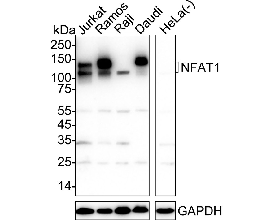
Western blot analysis of NFAT1 on different lysates with Rabbit anti-NFAT1 antibody at 1/1,000 dilution. Lane 1: Jurkat cell lysate, Lane 2: Ramos cell lysate, Lane 3: Raji cell lysate, Lane 4: Daudi cell lysate, Lane 5: HeLa cell lysate (negative), Lysates/proteins at 30 µg/Lane. Exposure time: 10 seconds; 4-20% SDS-PAGE gel. Proteins were transferred to a PVDF membrane and blocked with 5% NFDM/TBST for 1 hour at room temperature. The primary antibody at 1/1,000 dilution was used in 5% NFDM/TBST at 4℃ overnight. Goat Anti-Rabbit IgG - HRP Secondary Antibody at 1/50,000 dilution was used for 1 hour at room temperature.IHC
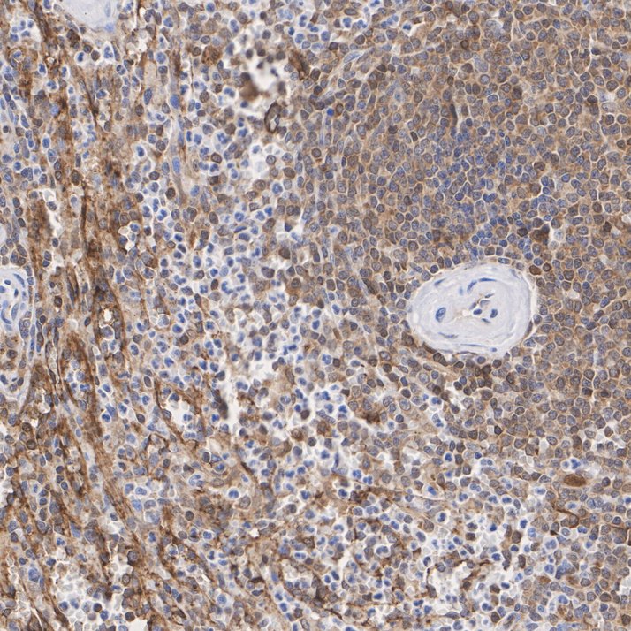
Immunohistochemical analysis of paraffin-embedded human spleen tissue with Rabbit anti-NFAT1 antibody at 1/1,000 dilution. The section was pre-treated using heat mediated antigen retrieval with sodium citrate buffer (pH 6.0) for 2 minutes. The tissues were blocked in 1% BSA for 20 minutes at room temperature, washed with ddH2O and PBS, and then probed with the primary antibody at 1/1,000 dilution for 1 hour at room temperature. The detection was performed using an HRP conjugated compact polymer system. DAB was used as the chromogen. Tissues were counterstained with hematoxylin and mounted with DPX.IHC
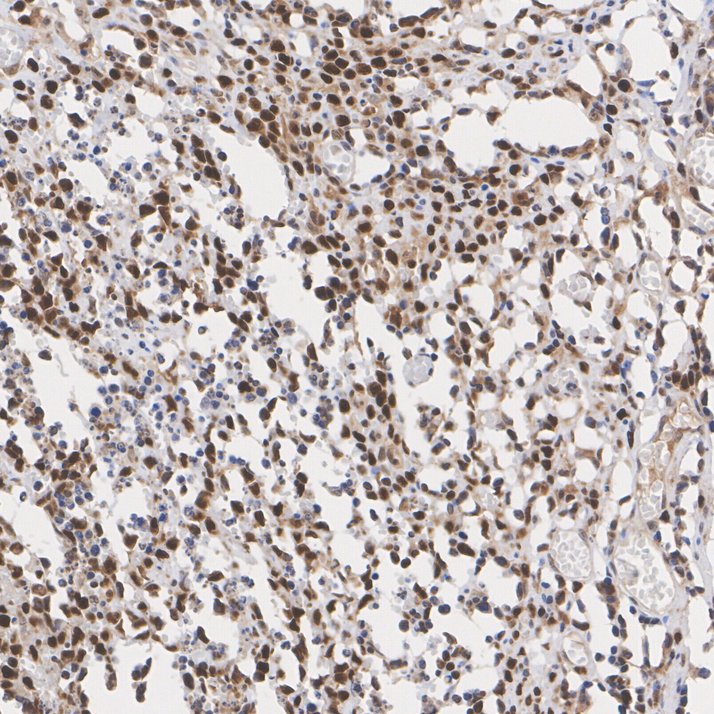
Immunohistochemical analysis of paraffin-embedded human NK-T-cell lymphoma tissue with Rabbit anti-NFAT1 antibody at 1/1,000 dilution. The section was pre-treated using heat mediated antigen retrieval with sodium citrate buffer (pH 6.0) for 2 minutes. The tissues were blocked in 1% BSA for 20 minutes at room temperature, washed with ddH2O and PBS, and then probed with the primary antibody at 1/1,000 dilution for 1 hour at room temperature. The detection was performed using an HRP conjugated compact polymer system. DAB was used as the chromogen. Tissues were counterstained with hematoxylin and mounted with DPX.IHC
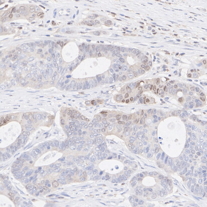
Immunohistochemical analysis of paraffin-embedded human colon cancer tissue with Rabbit anti-NFAT1 antibody at 1/1,000 dilution. The section was pre-treated using heat mediated antigen retrieval with sodium citrate buffer (pH 6.0) for 2 minutes. The tissues were blocked in 1% BSA for 20 minutes at room temperature, washed with ddH2O and PBS, and then probed with the primary antibody at 1/1,000 dilution for 1 hour at room temperature. The detection was performed using an HRP conjugated compact polymer system. DAB was used as the chromogen. Tissues were counterstained with hematoxylin and mounted with DPX.| Product Name | NFAT1 Recombinant Rabbit Monoclonal Antibody |
|---|---|
| Antibody Type | Primary Antibodies |
| Product description | The NFAT (nuclear factor of activated T cells) family of transcription factors regulates cytokine expression in T cells. Members of the family include NFATc1 (NFATc), NFATc2 (NFATp), NFATn, NFATc3 (NFAT4, NFATx) and NFATc4 (NFAT3). Recognition of antigen by the T cell receptor (TCR) eventually activates the calcium-dependent protein phosphatase calcineurin. Once activated, calcineurin stimulates the translocation of NFATc1 (cytoplasmic) from the cytoplasm to the nucleus where it associates with NFATn (nuclear). Like NFATc1, NFATc2 resides in the cytoplasm and translocates to the nucleus subsequent to activation of calcineurin. Once in the nucleus, NFATc2 synergizes with AP-1 transcription factors to initiate transcription of cytokine genes. NFATc3 and NFATc4 share 65% sequence identity with other members of the NFAT family. They are similar to NFATc2 in that they also synergize with the AP-1 family of proteins. |
| Immunogen | Synthetic peptide within Human NFAT1 aa 21-70 / 925. |
| Clonality | Monoclonal |
|---|---|
| Isotype | IgG |
| Host Species | Recombinant rabbit |
| Tested Applications | IHCWB |
| WB:1:1000 IHC:1:1000 |
|
| Species Reactivity | Human |
| Concentration | 1mg/ml |
| Purification | Protein A |
| Alternative Names | AI607462 antibody
cytoplasmic 2 antibody
KIAA0611 antibody
NF ATc2 antibody
NF ATp antibody
NF-ATc2 antibody
NF-ATp antibody
NFAC2_HUMAN antibody
NFAT 1 antibody
NFAT pre existing subunit antibody
NFAT pre-existing subunit antibody
NFAT transcription complex preexisting component antibody NFAT1 antibody NFAT1-D antibody NFATc2 antibody NFATp antibody Nuclear factor of activated T cells cytoplasmic 2 antibody Nuclear factor of activated T cells cytoplasmic calcineurin dependent 2 antibody Nuclear factor of activated T cells pre-existing component antibody Nuclear factor of activated T cells preexisting component antibody Nuclear factor of activated T-cells antibody Preexisting nuclear factor of activated T cells 2 antibody T cell transcription factor NFAT 1 antibody T-cell transcription factor NFAT1 antibody |
|---|---|
| Molecular Weight(MW) | 100kDa(Observed band size: 120/140kDa) |
| Cellular Localization | Cytoplasm. Nucleus. |
| SwissProt ID | Q13469 |
|---|
WB
Western blot analysis of NFAT1 on different lysates with Rabbit anti-NFAT1 antibody at 1/1,000 dilution. Lane 1: Jurkat cell lysate, Lane 2: Ramos cell lysate, Lane 3: Raji cell lysate, Lane 4: Daudi cell lysate, Lane 5: HeLa cell lysate (negative), Lysates/proteins at 30 µg/Lane. Exposure time: 10 seconds; 4-20% SDS-PAGE gel. Proteins were transferred to a PVDF membrane and blocked with 5% NFDM/TBST for 1 hour at room temperature. The primary antibody at 1/1,000 dilution was used in 5% NFDM/TBST at 4℃ overnight. Goat Anti-Rabbit IgG - HRP Secondary Antibody at 1/50,000 dilution was used for 1 hour at room temperature.
IHC
Immunohistochemical analysis of paraffin-embedded human spleen tissue with Rabbit anti-NFAT1 antibody at 1/1,000 dilution. The section was pre-treated using heat mediated antigen retrieval with sodium citrate buffer (pH 6.0) for 2 minutes. The tissues were blocked in 1% BSA for 20 minutes at room temperature, washed with ddH2O and PBS, and then probed with the primary antibody at 1/1,000 dilution for 1 hour at room temperature. The detection was performed using an HRP conjugated compact polymer system. DAB was used as the chromogen. Tissues were counterstained with hematoxylin and mounted with DPX.
IHC
Immunohistochemical analysis of paraffin-embedded human NK-T-cell lymphoma tissue with Rabbit anti-NFAT1 antibody at 1/1,000 dilution. The section was pre-treated using heat mediated antigen retrieval with sodium citrate buffer (pH 6.0) for 2 minutes. The tissues were blocked in 1% BSA for 20 minutes at room temperature, washed with ddH2O and PBS, and then probed with the primary antibody at 1/1,000 dilution for 1 hour at room temperature. The detection was performed using an HRP conjugated compact polymer system. DAB was used as the chromogen. Tissues were counterstained with hematoxylin and mounted with DPX.
IHC
Immunohistochemical analysis of paraffin-embedded human colon cancer tissue with Rabbit anti-NFAT1 antibody at 1/1,000 dilution. The section was pre-treated using heat mediated antigen retrieval with sodium citrate buffer (pH 6.0) for 2 minutes. The tissues were blocked in 1% BSA for 20 minutes at room temperature, washed with ddH2O and PBS, and then probed with the primary antibody at 1/1,000 dilution for 1 hour at room temperature. The detection was performed using an HRP conjugated compact polymer system. DAB was used as the chromogen. Tissues were counterstained with hematoxylin and mounted with DPX.| Application Notes | WB:1:1000 IHC:1:1000 |
|---|
| Form | Liquid |
|---|---|
| Storage Instructions | Store at +4℃ after thawing. Aliquot store at -20℃ or -80℃. Avoid repeated freeze / thaw cycles. |
| Storage Buffer | 1*TBS (pH7.4), 1%BSA, 40%Glycerol. Preservative: 0.05% Sodium Azide. |


