Application
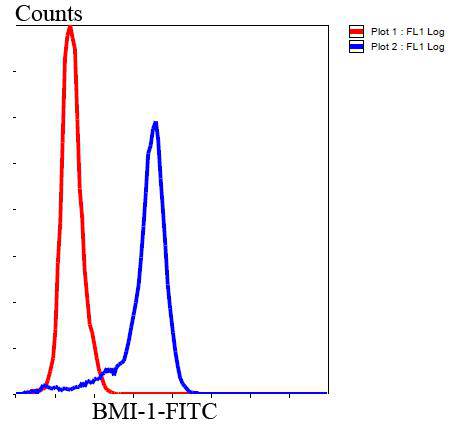
Fig8: Flow cytometric analysis of Hela cells with BMI1 antibody at 1/100 dilution (blue) compared with an unlabelled control (cells without incubation with primary antibody; red). Goat anti mouse IgG (FITC) was used as the secondary antibody.Application
Fig8: Flow cytometric analysis of Hela cells with BMI1 antibody at 1/100 dilution (blue) compared with an unlabelled control (cells without incubation with primary antibody; red). Goat anti mouse IgG (FITC) was used as the secondary antibody.Application
Fig8: Flow cytometric analysis of Hela cells with BMI1 antibody at 1/100 dilution (blue) compared with an unlabelled control (cells without incubation with primary antibody; red). Goat anti mouse IgG (FITC) was used as the secondary antibody.Application
Fig8: Flow cytometric analysis of Hela cells with BMI1 antibody at 1/100 dilution (blue) compared with an unlabelled control (cells without incubation with primary antibody; red). Goat anti mouse IgG (FITC) was used as the secondary antibody.Application
Fig8: Flow cytometric analysis of Hela cells with BMI1 antibody at 1/100 dilution (blue) compared with an unlabelled control (cells without incubation with primary antibody; red). Goat anti mouse IgG (FITC) was used as the secondary antibody.Application
Fig8: Flow cytometric analysis of Hela cells with BMI1 antibody at 1/100 dilution (blue) compared with an unlabelled control (cells without incubation with primary antibody; red). Goat anti mouse IgG (FITC) was used as the secondary antibody.Application
Fig8: Flow cytometric analysis of Hela cells with BMI1 antibody at 1/100 dilution (blue) compared with an unlabelled control (cells without incubation with primary antibody; red). Goat anti mouse IgG (FITC) was used as the secondary antibody.Application
Fig8: Flow cytometric analysis of Hela cells with BMI1 antibody at 1/100 dilution (blue) compared with an unlabelled control (cells without incubation with primary antibody; red). Goat anti mouse IgG (FITC) was used as the secondary antibody.| Product Name | Bmi1 [B3-G5] |
|---|---|
| Antibody Type | Primary Antibodies |
| Antigen Alias | B lymphoma Mo MLV insertion region (mouse) antibody B lymphoma Mo MLV insertion region 1 homolog antibody Bmi 1 antibody BMI1 antibody BMI1 polycomb ring finger oncogene antibody BMI1_HUMAN antibody Flvi 2/bmi 1 antibody FLVI2/BMI1 antibody MGC12685 antibody Murine leukemia viral (bmi 1) oncogene homolog antibody Oncogene BMI 1 antibody PCGF 4 antibody PCGF4 antibody Polycomb complex protein BMI 1 antibody Polycomb complex protein BMI-1 antibody Polycomb group protein Bmi1 antibody Polycomb group ring finger 4 antibody Polycomb group RING finger protein 4 antibody RING finger protein 51 antibody RNF 51 antibody RNF51 antibody |
| Product description | The Bmi-1 was identified initially as an oncogene that cooperates with c-myc in the generation of B-cell lymphoma. It contributes to the maintenance of cell identity, stem cell self-renewal, cell cycle regulation, and oncogenesis by maintaining the silenced state of genes that promote cell lineage specification, cell death, and cell-cycle arrest. |
| Immunogen | Recombinant protein within human Bmi1 full sequence. |
| Clonality | Monoclonal |
|---|---|
| Isotype | IgG2a |
| Host Species | Mouse |
| Tested Applications | WBICCIHCFC |
| WB:1:1,000 IHC:1:200 ICC:1:200 FC:1:100-1:200 |
|
| Species Reactivity | HumanMouseRat |
| Concentration | 2mg/ml |
| Alternative Names | B lymphoma Mo MLV insertion region (mouse) antibody B lymphoma Mo MLV insertion region 1 homolog antibody Bmi 1 antibody BMI1 antibody BMI1 polycomb ring finger oncogene antibody BMI1_HUMAN antibody Flvi 2/bmi 1 antibody FLVI2/BMI1 antibody MGC12685 antibody Murine leukemia viral (bmi 1) oncogene homolog antibody Oncogene BMI 1 antibody PCGF 4 antibody PCGF4 antibody Polycomb complex protein BMI 1 antibody Polycomb complex protein BMI-1 antibody Polycomb group protein Bmi1 antibody Polycomb group ring finger 4 antibody Polycomb group RING finger protein 4 antibody RING finger protein 51 antibody RNF 51 antibody RNF51 antibody |
|---|---|
| Molecular Weight(MW) | 37kDa |
| Cellular Localization | Nucleus, Cytoplasm |
Application
Fig8: Flow cytometric analysis of Hela cells with BMI1 antibody at 1/100 dilution (blue) compared with an unlabelled control (cells without incubation with primary antibody; red). Goat anti mouse IgG (FITC) was used as the secondary antibody.
Application
Fig8: Flow cytometric analysis of Hela cells with BMI1 antibody at 1/100 dilution (blue) compared with an unlabelled control (cells without incubation with primary antibody; red). Goat anti mouse IgG (FITC) was used as the secondary antibody.
Application
Fig8: Flow cytometric analysis of Hela cells with BMI1 antibody at 1/100 dilution (blue) compared with an unlabelled control (cells without incubation with primary antibody; red). Goat anti mouse IgG (FITC) was used as the secondary antibody.
Application
Fig8: Flow cytometric analysis of Hela cells with BMI1 antibody at 1/100 dilution (blue) compared with an unlabelled control (cells without incubation with primary antibody; red). Goat anti mouse IgG (FITC) was used as the secondary antibody.
Application
Fig8: Flow cytometric analysis of Hela cells with BMI1 antibody at 1/100 dilution (blue) compared with an unlabelled control (cells without incubation with primary antibody; red). Goat anti mouse IgG (FITC) was used as the secondary antibody.
Application
Fig8: Flow cytometric analysis of Hela cells with BMI1 antibody at 1/100 dilution (blue) compared with an unlabelled control (cells without incubation with primary antibody; red). Goat anti mouse IgG (FITC) was used as the secondary antibody.
Application
Fig8: Flow cytometric analysis of Hela cells with BMI1 antibody at 1/100 dilution (blue) compared with an unlabelled control (cells without incubation with primary antibody; red). Goat anti mouse IgG (FITC) was used as the secondary antibody.
Application
Fig8: Flow cytometric analysis of Hela cells with BMI1 antibody at 1/100 dilution (blue) compared with an unlabelled control (cells without incubation with primary antibody; red). Goat anti mouse IgG (FITC) was used as the secondary antibody.| Positive Control | Hela, HepG2, Jurkat, 293T, MCF-7, PC12, K562, NIH/3T3, A549, LOVO, human tonsil tissue, human colon cancer tissue, human breast cancer tissue, mouse kidney tissue, human kidney tissue, human brain tissue. |
|---|---|
| Application Notes | WB:1:1,000 IHC:1:200 ICC:1:200 FC:1:100-1:200 |
| Form | Liquid |
|---|---|
| Storage Instructions | Store at +4℃ after thawing. Aliquot store at -20℃. Avoid repeated freeze / thaw cycles. |
| Storage Buffer | 1*TBS (pH7.4), 1%BSA, 40%Glycerol. Preservative: 0.05% Sodium Azide. |


