Application
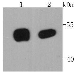
Fig1: Western blot analysis of p53 on different cell lysates using anti-p53 antibody at 1/1000 dilution. Positive control: Lane 1: 293T Lane 2: A431Application
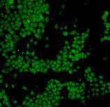
Fig2: ICC staining p53 in A431 cells (green). Cells were fixed in paraformaldehyde, permeabilised with 0.25% Triton X100/PBS.Application
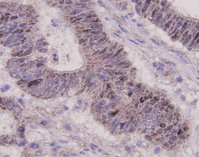
Fig3: Immunohistochemical analysis of paraffin-embedded human colonic carcinoma tissue using anti-p53 antibody. Counter stained with hematoxylin.Application
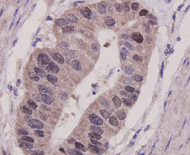
Fig4: Immunohistochemical analysis of paraffin-embedded human gastric carcinoma tissue using anti-p53 antibody. Counter stained with hematoxylin.Application
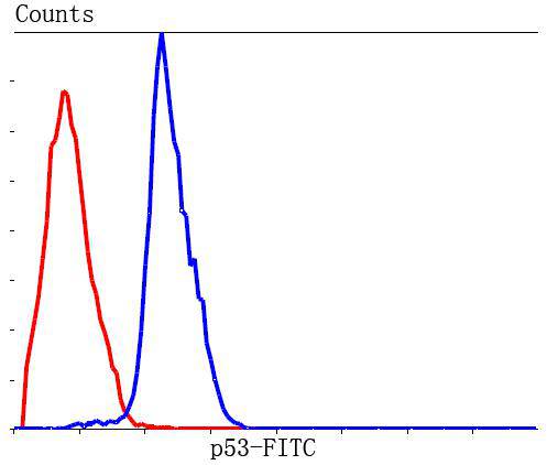
Fig5: Flow cytometric analysis of Hela cells with p53 antibody at 1/100 dilution (blue) compared with an unlabelled control (cells without incubation with primary antibody; red). Goat anti mouse IgG (FITC) was used as the secondary antibody.| Product Name | p53 [7-5] |
|---|---|
| Antibody Type | Primary Antibodies |
| Antigen Alias | Antigen NY-CO-13 antibody BCC7 antibody Cellular tumor antigen p53 antibody FLJ92943 antibody LFS1 antibody Mutant tumor protein 53 antibody p53 antibody p53 tumor suppressor antibody P53_HUMAN antibody Phosphoprotein p53 antibody Tp53 antibody Transformation related protein 53 antibody TRP53 antibody Tumor protein 53 antibody Tumor protein p53 antibody Tumor suppressor p53 antibody |
| Product description | p53 (also known as protein 53 or tumor protein 53), is a tumor suppressor protein that in humans is encoded by the TP53 gene. p53 is crucial in multicellular organisms, where it regulates the cell cycle and, thus, functions as a tumor suppressor that is involved in preventing cancer. As such, p53 has been described as "the guardian of the genome" because of its role in conserving stability by preventing genome mutation. |
| Immunogen | recombinant protein |
| Clonality | Monoclonal |
|---|---|
| Isotype | Ig2b |
| Host Species | Mouse |
| Tested Applications | WBICCIHCFC |
| WB:1:1,000 IHC:1:100-1:200 FC:1:100-1:200 ICC:1:100-1:200 |
|
| Species Reactivity | Human |
| Concentration | 2mg/ml |
| Alternative Names | Antigen NY-CO-13 antibody
BCC7 antibody Cellular tumor antigen p53 antibody FLJ92943 antibody LFS1 antibody Mutant tumor protein 53 antibody p53 antibody p53 tumor suppressor antibody P53_HUMAN antibody Phosphoprotein p53 antibody Tp53 antibody Transformation related protein 53 antibody TRP53 antibody Tumor protein 53 antibody Tumor protein p53 antibody Tumor suppressor p53 antibody |
|---|---|
| Molecular Weight(MW) | 53 kDa |
| Cellular Localization | Nucleus, Cytoplasm |
| SwissProt ID | P04637 |
|---|
Application
Fig1: Western blot analysis of p53 on different cell lysates using anti-p53 antibody at 1/1000 dilution. Positive control: Lane 1: 293T Lane 2: A431
Application
Fig2: ICC staining p53 in A431 cells (green). Cells were fixed in paraformaldehyde, permeabilised with 0.25% Triton X100/PBS.
Application
Fig3: Immunohistochemical analysis of paraffin-embedded human colonic carcinoma tissue using anti-p53 antibody. Counter stained with hematoxylin.
Application
Fig4: Immunohistochemical analysis of paraffin-embedded human gastric carcinoma tissue using anti-p53 antibody. Counter stained with hematoxylin.
Application
Fig5: Flow cytometric analysis of Hela cells with p53 antibody at 1/100 dilution (blue) compared with an unlabelled control (cells without incubation with primary antibody; red). Goat anti mouse IgG (FITC) was used as the secondary antibody.| Positive Control | A431, 293T, human colonic carcinoma, human gastric carcinoma. |
|---|---|
| Application Notes | WB:1:1,000 IHC:1:100-1:200 FC:1:100-1:200 ICC:1:100-1:200 |
| Form | Liquid |
|---|---|
| Storage Instructions | Store at +4℃ after thawing. Aliquot store at -20℃ or -80℃. Avoid repeated freeze / thaw cycles. |
| Storage Buffer | 1*TBS (pH7.4), 1%BSA, 40%Glycerol. Preservative: 0.05% Sodium Azide. |


