Application
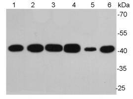
Fig1: Western blot analysis of Beta-Fig1: Western blot analysis of Beta-actin on different lysates using anti-Beta-actin antibody at 1/10,000 dilution.Positive control:Lane 1: NIH/3T3Lane 2: PC12Lane 3: MCF-7Lane 4: HepG2Lane 5: HelaLane 6: Mouse lungApplication
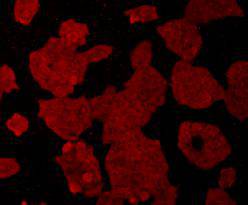
Fig2: ICC staining of Beta-actin in Hela cells (red). Cells were fixed in paraformaldehyde, permeabilised with 0.25% Triton X100/PBS.Application
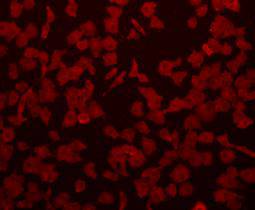
Fig3: ICC staining of Beta-actin in A549 cells (red). Cells were fixed in paraformaldehyde, permeabilised with 0.25% Triton X100/PBS.Application
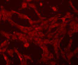
Fig4: ICC staining of Beta-actin in NIH-3T3 cells (red). Cells were fixed in paraformaldehyde, permeabilised with 0.25% Triton X100/PBS.Application
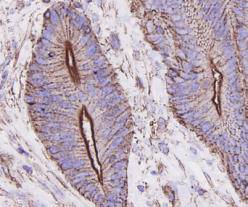
Fig5: Immunohistochemical analysis of paraffin-embedded human colon carcinoma tissue using anti-Beta-actin antibody. Counter stained with hematoxylin.Application
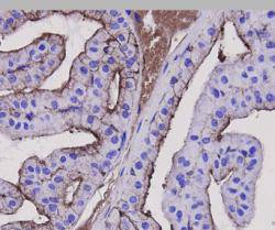
Fig6: Immunohistochemical analysis of paraffin-embedded mouse prostate tissue using anti-Beta-actin antibody. Counter stained with hematoxylin.Application
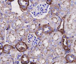
Fig7: Immunohistochemical analysis of paraffin-embedded mouse kidney tissue using anti-Beta-actin antibody. Counter stained with hematoxylin.Application
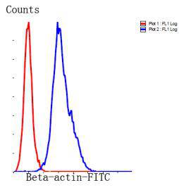
Fig8: Flow cytometric analysis of Hela?cells with Beta-actin antibody at 1/100 dilution (blue) compared with an unlabelled control (cells without incubation with primary antibody; red). Goat anti mouse IgG (FITC) was used as the secondary antibody.| Product Name | Beta-Actin [A2-F6] |
|---|---|
| Antibody Type | Primary Antibodies |
| Antigen Alias | A26C1A antibody A26C1B antibody ACTB antibody ACTB_HUMAN antibody Actin beta antibody Actin cytoplasmic 1 antibody Actin, cytoplasmic 1, N-terminally processed antibody Actx antibody b actin antibody Beta cytoskeletal actin antibody Beta-actin antibody BRWS1 antibody E430023M04Rik antibody MGC128179 antibody PS1TP5 binding protein 1 antibody PS1TP5BP1 antibody |
| Product description | Beta actin also know as Cytoplasmic Actin is a 42 kDa, highly conserved protein, ubiquitously expressed in all eukaryotic cells. They comprise, along with microtubules, a major component of the cytoskeleton. β-Actin is a relatively stable cytoskeletal protein normally at a constant level in cells, regardless of experimental treatment or technical procedure. For this reason, measurement of β -Actin is generally used as an internal control for experimental error. |
| Immunogen | Synthetic peptide (KLH-coupled) within human Beta-actin N terminal. |
| Clonality | Monoclonal |
|---|---|
| Isotype | IgG1 |
| Host Species | Mouse |
| Tested Applications | WBICCIHCFC |
| WB:1:10,000 ICC:1:100-1:200 IHC:1:100-1:200 FC:1:100-1:200 |
|
| Species Reactivity | HumanMouseRat |
| Concentration | 2mg/ml |
| Alternative Names | A26C1A antibody
A26C1B antibody
ACTB antibody
ACTB_HUMAN antibody
Actin beta antibody
Actin cytoplasmic 1 antibody
Actin cytoplasmic 1 N-terminally processed antibody Actx antibody b actin antibody Beta cytoskeletal actin antibody Beta-actin antibody BRWS1 antibody E430023M04Rik antibody MGC128179 antibody PS1TP5 binding protein 1 antibody PS1TP5BP1 antibody |
|---|---|
| Molecular Weight(MW) | 42kDa |
| Cellular Localization | Cytoplasm |
Application
Fig1: Western blot analysis of Beta-Fig1: Western blot analysis of Beta-actin on different lysates using anti-Beta-actin antibody at 1/10,000 dilution.Positive control:Lane 1: NIH/3T3Lane 2: PC12Lane 3: MCF-7Lane 4: HepG2Lane 5: HelaLane 6: Mouse lung
Application
Fig2: ICC staining of Beta-actin in Hela cells (red). Cells were fixed in paraformaldehyde, permeabilised with 0.25% Triton X100/PBS.
Application
Fig3: ICC staining of Beta-actin in A549 cells (red). Cells were fixed in paraformaldehyde, permeabilised with 0.25% Triton X100/PBS.
Application
Fig4: ICC staining of Beta-actin in NIH-3T3 cells (red). Cells were fixed in paraformaldehyde, permeabilised with 0.25% Triton X100/PBS.
Application
Fig5: Immunohistochemical analysis of paraffin-embedded human colon carcinoma tissue using anti-Beta-actin antibody. Counter stained with hematoxylin.
Application
Fig6: Immunohistochemical analysis of paraffin-embedded mouse prostate tissue using anti-Beta-actin antibody. Counter stained with hematoxylin.
Application
Fig7: Immunohistochemical analysis of paraffin-embedded mouse kidney tissue using anti-Beta-actin antibody. Counter stained with hematoxylin.
Application
Fig8: Flow cytometric analysis of Hela?cells with Beta-actin antibody at 1/100 dilution (blue) compared with an unlabelled control (cells without incubation with primary antibody; red). Goat anti mouse IgG (FITC) was used as the secondary antibody.| Positive Control | NIH/3T3, PC12, MCF-7, HepG2, Hela, mouse lung A549, human colon carcinoma tissue, mouse prostate tissue , mouse kidney tissue |
|---|---|
| Application Notes | WB:1:10,000 ICC:1:100-1:200 IHC:1:100-1:200 FC:1:100-1:200 |
| Form | Liquid |
|---|---|
| Storage Instructions | Store at +4℃ after thawing. Aliquot store at -20℃ or -80℃. Avoid repeated freeze / thaw cycles. |
| Storage Buffer | 1*TBS (pH7.4), 1%BSA, 40%Glycerol. Preservative: 0.05% Sodium Azide. |


