Application
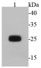
Fig1: Western blot analysis of GFP on transfected K562 with GFP using anti-GFP antibody at 1/2000 dilution.Application
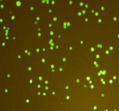
Fig2: ICC staining GFP in transfected K562 cells (green). Cells were fixed in paraformaldehyde, permeabilised with 0.25% Triton X100/PBS.| Product Name | GFP [2-D8] |
|---|---|
| Antibody Type | Primary Antibodies |
| Antigen Alias | GFP antibody Green fluorescent protein antibody yfp antibody |
| Product description | Green fluorescence protein (GFP) is derived from the jellyfish Aequorea victoria, which emits green light (emission peak at a wavelength of 509 nm) when excited by blue light (excitation peak at a wavelength of 395 nm). GFP fluorescence is stable under fixation conditions and suitable for a variety of applications. It has been widely used as a reporter for gene expression, enabling researchers to visualize and localize GFP-tagged proteins within living cells without chemical staining. |
| Immunogen | recombinant protein |
| Clonality | Monoclonal |
|---|---|
| Isotype | IgG1 |
| Host Species | Mouse |
| Tested Applications | WBICCICC/IFIPFC |
| WB:1:2,000 ICC:1:200 |
|
| Concentration | 2mg/ml |
| Alternative Names | GFP antibody Green fluorescent protein antibody yfp antibody |
|---|---|
| Molecular Weight(MW) | 26 kDa |
| SwissProt ID | P42212 |
|---|
Application
Fig1: Western blot analysis of GFP on transfected K562 with GFP using anti-GFP antibody at 1/2000 dilution.
Application
Fig2: ICC staining GFP in transfected K562 cells (green). Cells were fixed in paraformaldehyde, permeabilised with 0.25% Triton X100/PBS.| Application Notes | WB:1:2,000 ICC:1:200 |
|---|
| Form | Liquid |
|---|---|
| Storage Instructions | Store at +4℃ after thawing. Aliquot store at -20℃ or -80℃. Avoid repeated freeze / thaw cycles. |
| Storage Buffer | 1*TBS (pH7.4), 1%BSA, 40%Glycerol. Preservative: 0.05% Sodium Azide. |


