Application
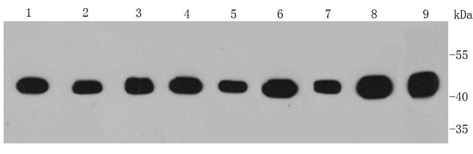
Fig1: Western blot analysis of IDH1 on different cell lysates using anti-IDH1 antibody at 1/1000 dilution. Positive control: Lane 1: Hela Lane 2: HepG2 Lane 3: A431 Lane 4: MCF-7 Lane 5: A549 Lane 6: Jurkat Lane 7: Human kidney Lane 8: Human brain Lane 9: Human liverApplication
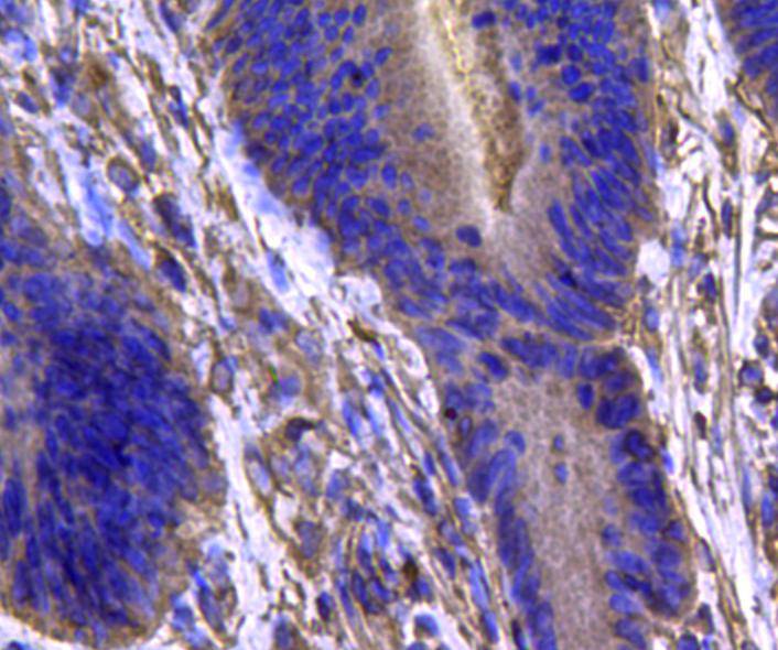
Fig2: Immunohistochemical analysis of paraffin-embedded human colon carcinoma tissue using anti-IDH1 antibody. Counter stained with hematoxylin.Application
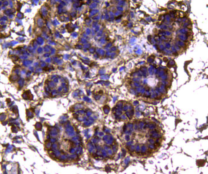
Fig3: Immunohistochemical analysis of paraffin-embedded human breast tissue using anti-IDH1 antibody. Counter stained with hematoxylin.Application
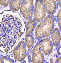
Fig4: Immunohistochemical analysis of paraffin-embedded human kidney tissue using anti-IDH1 antibody. Counter stained with hematoxylin.| Product Name | Isocitrate dehydrogenase/IDH1 [A11-B4] |
|---|---|
| Antibody Type | Primary Antibodies |
| Antigen Alias | Cytosolic NADP isocitrate dehydrogenase antibody Cytosolic NADP-isocitrate dehydrogenase antibody Epididymis luminal protein 216 antibody Epididymis secretory protein Li 26 antibody HEL-216 antibody HEL-S-26 antibody ICDH antibody IDCD antibody IDH antibody IDH1 antibody IDHC_HUMAN antibody IDP antibody IDPC antibody Isocitrate dehydrogenase [NADP] cytoplasmic antibody Isocitrate dehydrogenase 1 (NADP+) soluble antibody NADP dependent isocitrate dehydrogenase cytosolic antibody NADP dependent isocitrate dehydrogenase peroxisomal antibody NADP(+)-specific ICDH antibody Oxalosuccinate decarboxylase antibody PICD antibody |
| Product description | Isocitrate dehydrogenase (IDH) is an enzyme that catalyzes the oxidative decarboxylation of isocitrate, producing alpha-ketoglutarate (α-ketoglutarate) and CO2. In humans, IDH exists in three isoforms: IDH3 catalyzes the third step of the citric acid cycle while converting NAD+ to NADH in the mitochondria. The isoforms IDH1 and IDH2 catalyze the same reaction outside the context of the citric acid cycle and use NADP+ as a cofactor instead of NAD+. They localize to the cytosol as well as the mitochondrion and peroxisome. Mutations in IDH1 are also implicated in cancer. Originally mutations in IDH1 were detected in an integrated genomic analysis of human glioblastoma multiforme. In addition to being mutated in diffuse gliomas, IDH1 has also been shown to harbor mutations in human acute myeloid leukemia (AML). |
| Immunogen | recombinant protein |
| Clonality | Monoclonal |
|---|---|
| Isotype | IgG1 |
| Host Species | Mouse |
| Tested Applications | WBIHC |
| WB:1:1,000 IHC:1:100-1:200 |
|
| Species Reactivity | HumanZebra Fish |
| Concentration | 2mg/ml |
| Alternative Names | Cytosolic NADP isocitrate dehydrogenase antibody Cytosolic NADP-isocitrate dehydrogenase antibody Epididymis luminal protein 216 antibody Epididymis secretory protein Li 26 antibody HEL-216 antibody HEL-S-26 antibody ICDH antibody IDCD antibody IDH antibody IDH1 antibody IDHC_HUMAN antibody IDP antibody IDPC antibody Isocitrate dehydrogenase [NADP] cytoplasmic antibody Isocitrate dehydrogenase 1 (NADP+) soluble antibody NADP dependent isocitrate dehydrogenase cytosolic antibody NADP dependent isocitrate dehydrogenase peroxisomal antibody NADP(+)-specific ICDH antibody Oxalosuccinate decarboxylase antibody PICD antibody |
|---|---|
| Molecular Weight(MW) | 47 kDa |
| Cellular Localization | Cytoplasm |
| SwissProt ID | O75874 |
|---|
Application
Fig1: Western blot analysis of IDH1 on different cell lysates using anti-IDH1 antibody at 1/1000 dilution. Positive control: Lane 1: Hela Lane 2: HepG2 Lane 3: A431 Lane 4: MCF-7 Lane 5: A549 Lane 6: Jurkat Lane 7: Human kidney Lane 8: Human brain Lane 9: Human liver
Application
Fig2: Immunohistochemical analysis of paraffin-embedded human colon carcinoma tissue using anti-IDH1 antibody. Counter stained with hematoxylin.
Application
Fig3: Immunohistochemical analysis of paraffin-embedded human breast tissue using anti-IDH1 antibody. Counter stained with hematoxylin.
Application
Fig4: Immunohistochemical analysis of paraffin-embedded human kidney tissue using anti-IDH1 antibody. Counter stained with hematoxylin.| Positive Control | MCF-7, HepG2, Hela, A549, A431, Jurkat, human brain tissue, human liver tissue , human kidney tissue,human colon carcinoma tissue,human breast tissue. |
|---|---|
| Application Notes | WB:1:1,000 IHC:1:100-1:200 |
| Form | Liquid |
|---|---|
| Storage Instructions | Store at +4℃ after thawing. Aliquot store at -20℃ or -80℃. Avoid repeated freeze / thaw cycles. |
| Storage Buffer | 1*TBS (pH7.4), 1%BSA, 40%Glycerol. Preservative: 0.05% Sodium Azide. |


