Application
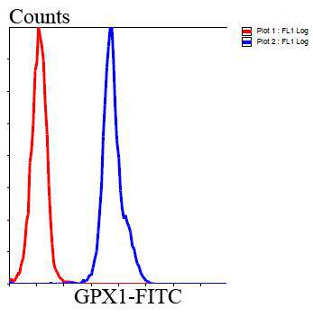
Fig5: Flow cytometric analysis of HepG2 cells with GPX1 antibody at 1/100 dilution (blue) compared with an unlabelled control (cells without incubation with primary antibody; red). Goat anti mouse IgG (FITC) was used as the secondary antibody.Application
Fig5: Flow cytometric analysis of HepG2 cells with GPX1 antibody at 1/100 dilution (blue) compared with an unlabelled control (cells without incubation with primary antibody; red). Goat anti mouse IgG (FITC) was used as the secondary antibody.Application
Fig5: Flow cytometric analysis of HepG2 cells with GPX1 antibody at 1/100 dilution (blue) compared with an unlabelled control (cells without incubation with primary antibody; red). Goat anti mouse IgG (FITC) was used as the secondary antibody.Application
Fig5: Flow cytometric analysis of HepG2 cells with GPX1 antibody at 1/100 dilution (blue) compared with an unlabelled control (cells without incubation with primary antibody; red). Goat anti mouse IgG (FITC) was used as the secondary antibody.Application
Fig5: Flow cytometric analysis of HepG2 cells with GPX1 antibody at 1/100 dilution (blue) compared with an unlabelled control (cells without incubation with primary antibody; red). Goat anti mouse IgG (FITC) was used as the secondary antibody.| Product Name | Glutathione Peroxidase 1 [C5-F6] |
|---|---|
| Antibody Type | Primary Antibodies |
| Antigen Alias | AL033363 antibody Cellular glutathione peroxidase antibody Glutathione peroxidase 1 antibody Glutathione peroxidase antibody GPx 1 antibody GPx-1 antibody GPX1 antibody GPX1_HUMAN antibody GPXD antibody GSHPx-1 antibody GSHPX1 antibody MGC14399 antibody MGC88245 antibody |
| Product description | GPX1 is ubiquitously expressed in many tissues, where it protects cells from oxidative stress. Within cells, it localizes to the cytoplasm and mitochondria. As a glutathione peroxidase, GPx1 functions in the detoxification of hydrogen peroxide, specifically by catalyzing the reduction of hydrogen peroxide to water. The glutathione peroxidase also catalyzes the reduction of other organic hydroperoxides, such as lipid peroxides, to the corresponding alcohols. GPx1 typically uses glutathione (GSH) as the reductant, but when glutathione synthetase (GSS) is, as in brain mitochondria, γ-glutamylcysteine can serve as the reductant instead. The protein encoded by this gene protects from CD95-induced apoptosis in cultured breast cancer cells and inhibits 5-lipoxygenase in blood cells, and its overexpression delays endothelial cell death and increases resistance to toxic challenges, especially oxidative stress. This protein is one of only a few proteins known in higher vertebrates to contain selenocysteine, which occurs at the active site of glutathione peroxidase and is coded by the nonsense (stop) codon TGA. |
| Immunogen | recombinant protein |
| Clonality | Monoclonal |
|---|---|
| Isotype | IgG2b |
| Host Species | Mouse |
| Tested Applications | WBICCIHCFC |
| WB:1:1,000 IHC:1:200 ICC:1:200 FC:1:100-1:200 |
|
| Species Reactivity | Human |
| Concentration | 2mg/ml |
| Alternative Names | AL033363 antibody Cellular glutathione peroxidase antibody Glutathione peroxidase 1 antibody Glutathione peroxidase antibody GPx 1 antibody GPx-1 antibody GPX1 antibody GPX1_HUMAN antibody GPXD antibody GSHPx-1 antibody GSHPX1 antibody MGC14399 antibody MGC88245 antibody |
|---|---|
| Molecular Weight(MW) | 22 kDa |
| Cellular Localization | Cytoplasm. |
| SwissProt ID | P07203 |
|---|
Application
Fig5: Flow cytometric analysis of HepG2 cells with GPX1 antibody at 1/100 dilution (blue) compared with an unlabelled control (cells without incubation with primary antibody; red). Goat anti mouse IgG (FITC) was used as the secondary antibody.
Application
Fig5: Flow cytometric analysis of HepG2 cells with GPX1 antibody at 1/100 dilution (blue) compared with an unlabelled control (cells without incubation with primary antibody; red). Goat anti mouse IgG (FITC) was used as the secondary antibody.
Application
Fig5: Flow cytometric analysis of HepG2 cells with GPX1 antibody at 1/100 dilution (blue) compared with an unlabelled control (cells without incubation with primary antibody; red). Goat anti mouse IgG (FITC) was used as the secondary antibody.
Application
Fig5: Flow cytometric analysis of HepG2 cells with GPX1 antibody at 1/100 dilution (blue) compared with an unlabelled control (cells without incubation with primary antibody; red). Goat anti mouse IgG (FITC) was used as the secondary antibody.
Application
Fig5: Flow cytometric analysis of HepG2 cells with GPX1 antibody at 1/100 dilution (blue) compared with an unlabelled control (cells without incubation with primary antibody; red). Goat anti mouse IgG (FITC) was used as the secondary antibody.| Positive Control | Hela, HepG2, 293T, human kidney tissue. |
|---|---|
| Application Notes | WB:1:1,000 IHC:1:200 ICC:1:200 FC:1:100-1:200 |
| Form | Liquid |
|---|---|
| Storage Instructions | Store at +4℃ after thawing. Aliquot store at -20℃ or -80℃. Avoid repeated freeze / thaw cycles. |
| Storage Buffer | 1*TBS (pH7.4), 1%BSA, 40%Glycerol. Preservative: 0.05% Sodium Azide. |


