Application
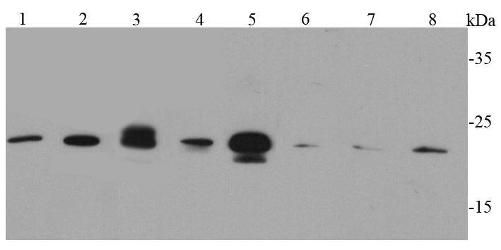
Fig1: Western blot analysis of Caveolin-1 on different cell lysates using anti-Caveolin-1 antibody at 1/500 dilution.Application
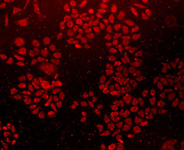
Fig2: ICC staining Caveolin-1 in L929 cells (red). Cells were fixed in paraformaldehyde, permeabilised with 0.25% Triton X100/PBS.Application
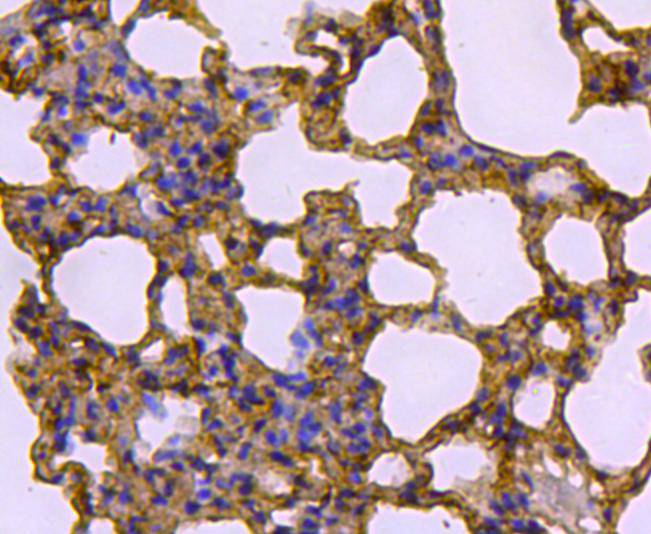
Fig3: Immunohistochemical analysis of paraffin-embedded rat lung tissue using anti-Caveolin-1 antibody. Counter stained with hematoxylin.Application
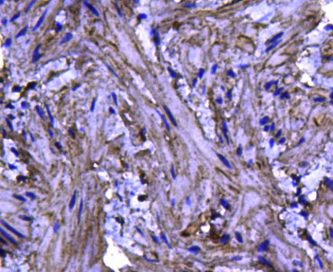
Fig4: Immunohistochemical analysis of paraffin-embedded mouse bladder tissue using anti-Caveolin-1 antibody. Counter stained with hematoxylin.Application
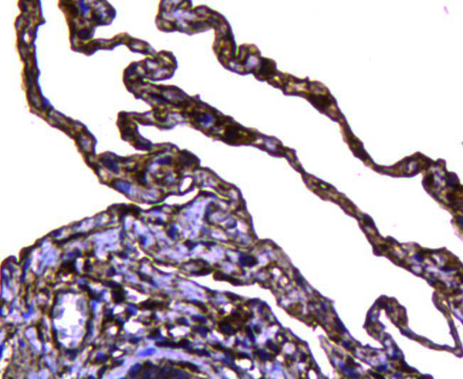
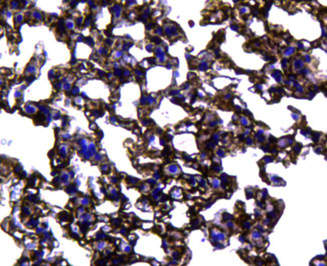
Fig5: Immunohistochemical analysis of paraffin-embedded human lung tissue using anti-Caveolin-1 antibody. Counter stained with hematoxylin.Application
Fig6: Immunohistochemical analysis of paraffin-embedded mouse lung tissue using anti-Caveolin-1 antibody. Counter stained with hematoxylin.| Product Name | Caveolin-1 [19-D6] |
|---|---|
| Antibody Type | Primary Antibodies |
| Antigen Alias | BSCL3 antibody CAV antibody CAV1 antibody CAV1_HUMAN antibody caveolae protein, 22 kD antibody caveolin 1 alpha isoform antibody caveolin 1 beta isoform antibody Caveolin 1 caveolae protein 22kDa antibody Caveolin-1 antibody Caveolin1 antibody cell growth-inhibiting protein 32 antibody CGL3 antibody LCCNS antibody MSTP085 antibody OTTHUMP00000025031 antibody PPH3 antibody VIP 21 antibody VIP21 antibody |
| Product description | Caveolin, an integral membrane protein, is a principal component of caveolae membranes in vivo. Two isoforms of caveolin have been identified: a slower migrating 24-kDa species (-isoform) and a faster migrating 21-kDa species (-isoform). Caveolins interact with multiple signaling molecules, such as the G-protein alpha subunit, tyrosine kinase receptors, PKCs, Src family tyrosine kinases and eNOS. Caveolin-1 has been implicated in the pathogenesis of mammary epithelial cell hyperplasia. |
| Immunogen | peptide |
| Clonality | Monoclonal |
|---|---|
| Isotype | IgG1 |
| Host Species | Mouse |
| Tested Applications | WBICCIHC |
| WB:1:500 IHC:1:200 ICC:1:200 |
|
| Species Reactivity | HumanMouseRat |
| Concentration | 2mg/ml |
| Alternative Names | BSCL3 antibody
CAV antibody
CAV1 antibody
CAV1_HUMAN antibody
caveolae protein 22 kD antibody caveolin 1 alpha isoform antibody caveolin 1 beta isoform antibody Caveolin 1 caveolae protein 22kDa antibody Caveolin-1 antibody Caveolin1 antibody cell growth-inhibiting protein 32 antibody CGL3 antibody LCCNS antibody MSTP085 antibody OTTHUMP00000025031 antibody PPH3 antibody VIP 21 antibody VIP21 antibody |
|---|---|
| Molecular Weight(MW) | 21 kDa |
| Cellular Localization | Cell membrane |
| SwissProt ID | Q03135 |
|---|
Application
Fig1: Western blot analysis of Caveolin-1 on different cell lysates using anti-Caveolin-1 antibody at 1/500 dilution.
Application
Fig2: ICC staining Caveolin-1 in L929 cells (red). Cells were fixed in paraformaldehyde, permeabilised with 0.25% Triton X100/PBS.
Application
Fig3: Immunohistochemical analysis of paraffin-embedded rat lung tissue using anti-Caveolin-1 antibody. Counter stained with hematoxylin.
Application
Fig4: Immunohistochemical analysis of paraffin-embedded mouse bladder tissue using anti-Caveolin-1 antibody. Counter stained with hematoxylin.
Application
Fig5: Immunohistochemical analysis of paraffin-embedded human lung tissue using anti-Caveolin-1 antibody. Counter stained with hematoxylin.
Application
Fig6: Immunohistochemical analysis of paraffin-embedded mouse lung tissue using anti-Caveolin-1 antibody. Counter stained with hematoxylin.| Positive Control | Hela, HepG2, A549, Jurkat, F9, L929, human lung tissue, mouse lung tissue, mouse liver tissue, rat lung tissue, mouse bladder tissue |
|---|---|
| Application Notes | WB:1:500 IHC:1:200 ICC:1:200 |
| Form | Liquid |
|---|---|
| Storage Instructions | Store at +4℃ after thawing. Aliquot store at -20℃ or -80℃. Avoid repeated freeze / thaw cycles. |
| Storage Buffer | 1*TBS (pH7.4), 1%BSA, 40%Glycerol. Preservative: 0.05% Sodium Azide. |


