Application
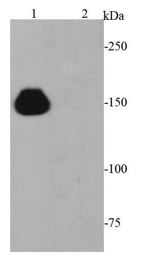
Fig1: Western blot analysis of CRISPR-Cas9 on 293T cell lysates using anti-CRISPR-Cas9 antibody at 1/5000 dilution. Lane 1: CRISPR-Cas9 transfected 293 cells lysate Lane 2: Non-transfected 293 cells lysateApplication
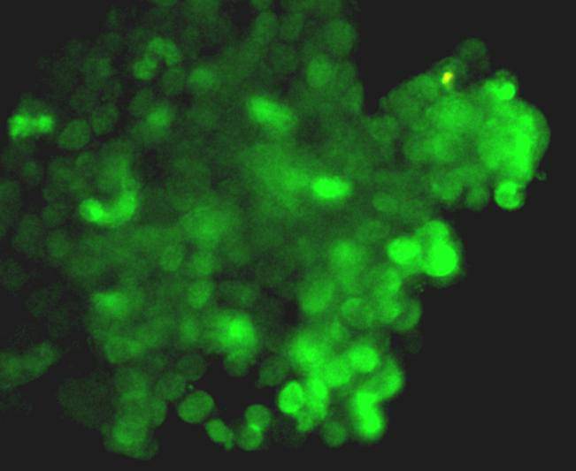
Fig2: Immunofluorescence staining CRISPR-Cas9 in CRISPR-Cas9 transfected 293T cells (green). Cells were fixed in paraformaldehyde, permeabilised with 0.25% Triton X100/PBS.| Product Name | CRISPR-Cas9 [8-A6-6] |
|---|---|
| Antibody Type | Primary Antibodies |
| Antigen Alias | Cas9 antibody CRISPR-associated endonuclease Cas9/Csn1 antibody CRISPR-Cas9/Csn1 antibody csn1 antibody SpyCas9 antibody |
| Product description | The Clustered Regularly Interspaced Short Palindromic Repeats (CRISPR) Type II system is currently the most commonly used RNA-Guided Endonuclease technology for genome engineering. There are two distinct components to this system: (1) a guide RNA and (2) an endonuclease, in this case the CRISPR associated (Cas) nuclease, Cas9. The gRNA/Cas9 complex is recruited to the target sequence by the base-pairing between the gRNA sequence and the complement to the target sequence in the genomic DNA. For successful binding of Cas9, the genomic target sequence must also contain the correct Protospacer Adjacent Motiff (PAM) sequence immediately following the target sequence. |
| Immunogen | recombinant protein |
| Clonality | Monoclonal |
|---|---|
| Isotype | IgM |
| Host Species | Mouse |
| Tested Applications | WBICC |
| WB:1:5,000 ICC:1:200 |
|
| Concentration | 2mg/ml |
| Alternative Names | Cas9 antibody CRISPR-associated endonuclease Cas9/Csn1 antibody CRISPR-Cas9/Csn1 antibody csn1 antibody SpyCas9 antibody |
|---|
Application
Fig1: Western blot analysis of CRISPR-Cas9 on 293T cell lysates using anti-CRISPR-Cas9 antibody at 1/5000 dilution. Lane 1: CRISPR-Cas9 transfected 293 cells lysate Lane 2: Non-transfected 293 cells lysate
Application
Fig2: Immunofluorescence staining CRISPR-Cas9 in CRISPR-Cas9 transfected 293T cells (green). Cells were fixed in paraformaldehyde, permeabilised with 0.25% Triton X100/PBS.| Positive Control | Human 293T cells transfected with CRISPR-Cas9 lysate. |
|---|---|
| Application Notes | WB:1:5,000 ICC:1:200 |
| Form | Liquid |
|---|---|
| Storage Instructions | Store at +4℃ after thawing. Aliquot store at -20℃ or -80℃. Avoid repeated freeze / thaw cycles. |
| Storage Buffer | 1*TBS (pH7.4), 1%BSA, 40%Glycerol. Preservative: 0.05% Sodium Azide. |


