WB
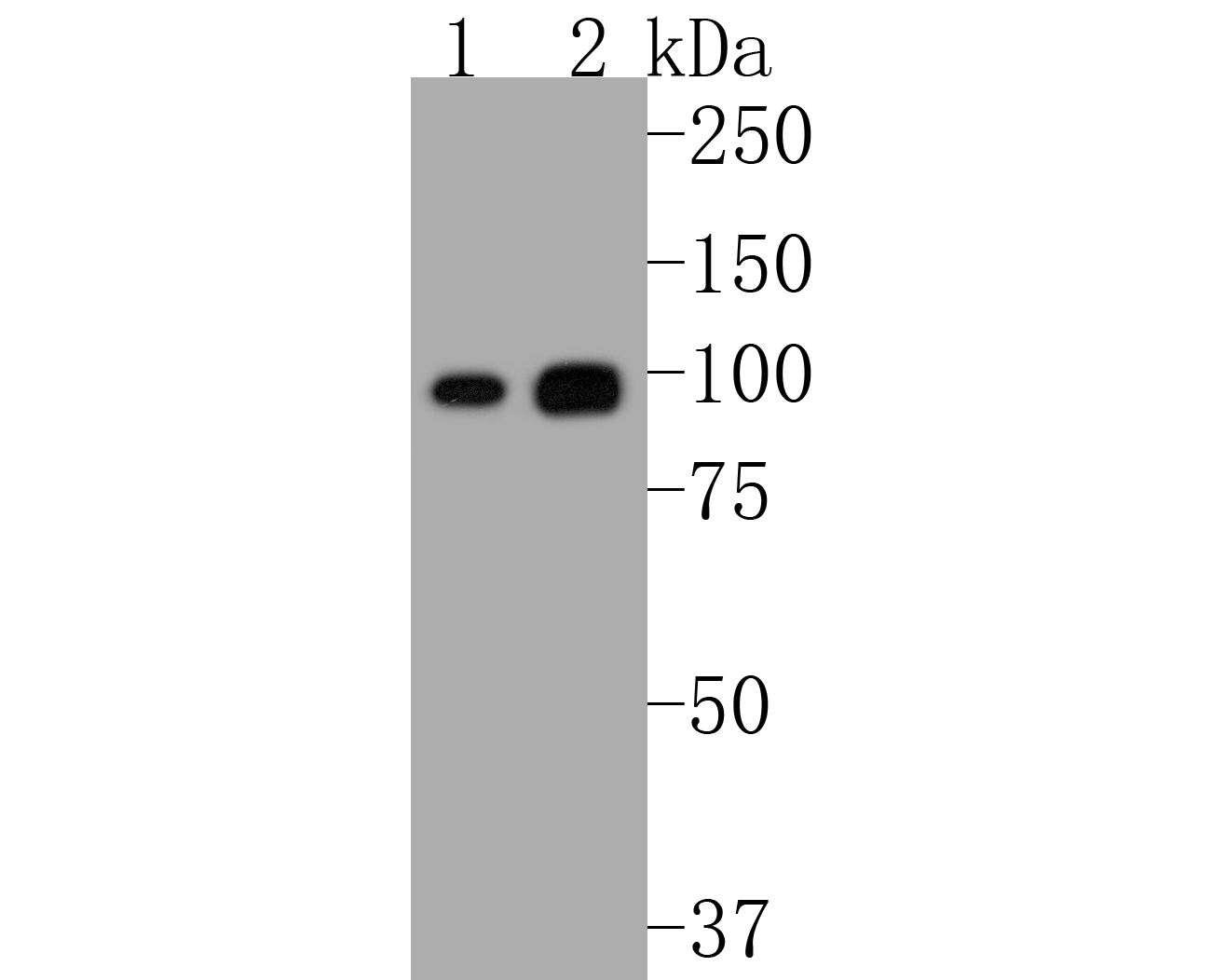
Western blot analysis of Phospho-Nrf2 (S40) on different lysates with Rabbit anti-Phospho-Nrf2 (S40) antibody at 1/2,000 dilution. Lane 1: HepG2 cell lysate Lane 2: Raji cell lysate Lysates/proteins at 10 µg/Lane. Exposure time: 30 seconds; 8% SDS-PAGE gel. Proteins were transferred to a PVDF membrane and blocked with 5% NFDM/TBST for 1 hour at room temperature. The primary antibody at 1/2,000 dilution was used in 5% NFDM/TBST at room temperature for 2 hours. Goat Anti-Rabbit IgG - HRP Secondary Antibody at 1/50,000 dilution was used for 1 hour at room temperature.ICC/IF
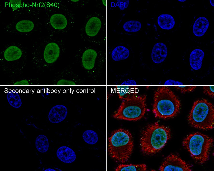
Immunocytochemistry analysis of HepG2 cells labeling Phospho-Nrf2 (S40) with Rabbit anti-Phospho-Nrf2 (S40) antibody at 1/250 dilution. Cells were fixed in 4% paraformaldehyde for 20 minutes at room temperature, permeabilized with 0.1% Triton X-100 in PBS for 5 minutes at room temperature, then blocked with 1% BSA in 10% negative goat serum for 1 hour at room temperature. Cells were then incubated with Rabbit anti-Phospho-Nrf2 (S40) antibody at 1/250 dilution in 1% BSA in PBST overnight at 4 ℃. Goat Anti-Rabbit IgG H&L (488) was used as the secondary antibody at 1/1,000 dilution. PBS instead of the primary antibody was used as the secondary antibody only control. Nuclear DNA was labelled in blue with DAPI. Beta tubulin (red) was stained at 1/100 dilution overnight at +4℃. Goat Anti-Mouse IgG H&L (594) was used as the secondary antibody at 1/1,000 dilution.IHC
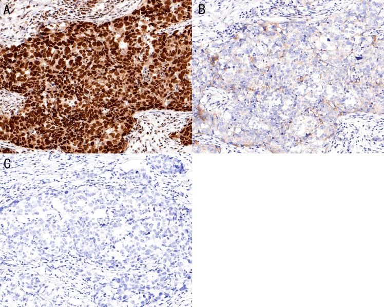
Immunohistochemical analysis of paraffin-embedded human breast carcinoma tissue using anti-Phospho-Nrf2 (S40) antibody. The section was pre-treated using heat mediated antigen retrieval with 10 mM sodium citrate buffer (pH 6.0). The section were untreated (A, C) and treated (B) with λ-PPase at 1/25 dilution at 30℃ for 30 minutes after antigen retrieval. The tissues were blocked in 1% BSA for 30 minutes at room temperature, washed with ddH2O and PBS, and then probed with the primary antibody (1/200) for 30 minutes at room temperature. Goat anti-Rabbit IgG-HRP UltraPolymer antibody was used for 20 minutes at room temperature. DAB was used as the chromogen. The section were counterstained with hematoxylin and mounted with DPX. PBS was used instead of the primary antibody as the negative control, and is shown in the inset (C).IHC
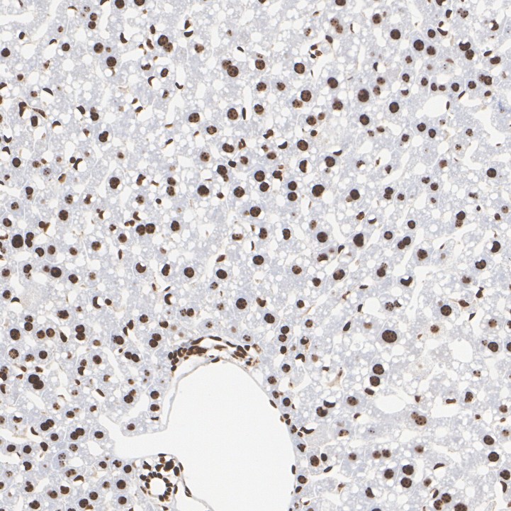
Immunohistochemical analysis of paraffin-embedded mouse liver tissue with Rabbit anti-Phospho-Nrf2 (S40) antibody at 1/1,000 dilution. The section was pre-treated using heat mediated antigen retrieval with sodium citrate buffer (pH 6.0) (high pressure) for 2 minutes. The tissues were blocked in 1% BSA for 20 minutes at room temperature, washed with ddH2O and PBS, and then probed with the primary antibody at 1/1,000 dilution for 1 hour at room temperature. The detection was performed using an HRP conjugated compact polymer system. DAB was used as the chromogen. Tissues were counterstained with hematoxylin and mounted with DPX.IHC
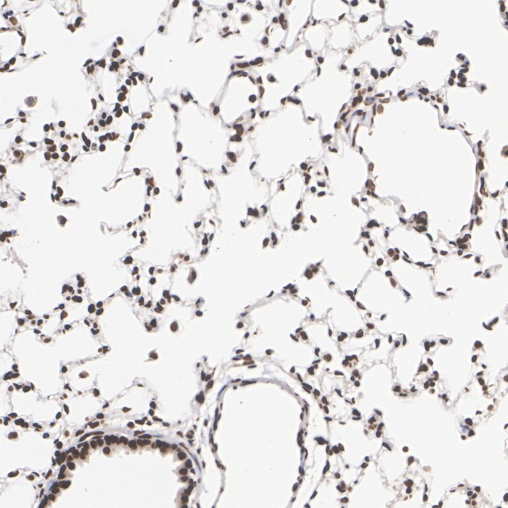
Immunohistochemical analysis of paraffin-embedded rat lung tissue with Rabbit anti-Phospho-Nrf2 (S40) antibody at 1/1,000 dilution. The section was pre-treated using heat mediated antigen retrieval with sodium citrate buffer (pH 6.0) (high pressure) for 2 minutes. The tissues were blocked in 1% BSA for 20 minutes at room temperature, washed with ddH2O and PBS, and then probed with the primary antibody at 1/1,000 dilution for 1 hour at room temperature. The detection was performed using an HRP conjugated compact polymer system. DAB was used as the chromogen. Tissues were counterstained with hematoxylin and mounted with DPX.FC
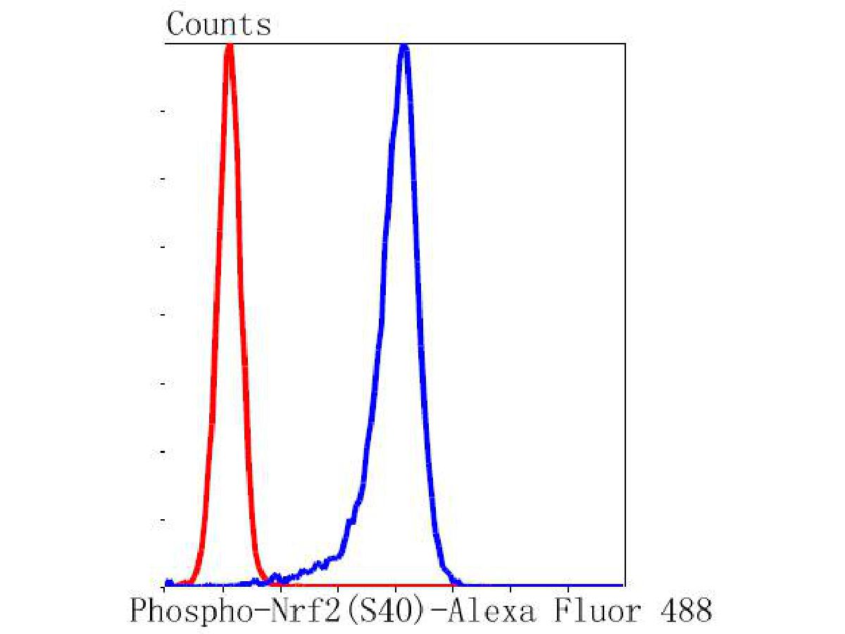
Flow cytometric analysis of Phospho-Nrf2 (S40) was done on K562 cells. The cells were fixed, permeabilized and stained with the primary antibody (1/50) (blue). After incubation of the primary antibody at room temperature for an hour, the cells were stained with a Alexa Fluor®488 conjugate-Goat anti-Rabbit IgG Secondary antibody at 1/1,000 dilution for 30 minutes.Unlabelled sample was used as a control (cells without incubation with primary antibody; red).| Product Name | Phospho-Nrf2 (S40) Recombinant Rabbit Monoclonal Antibody |
|---|---|
| Antibody Type | Primary Antibodies |
| Product description | The NF-E2 DNA binding protein is composed of two subunits, p45 and MafK. It regulates expression of globin genes in developing erythroid cells through interaction with Maf recognition elements (Mares). A family of NF-E2- related proteins, which are collectively known as the Cap ‘n’ collar (CNC) family and include Nrf1 (also designated TCF11), Nrf2 and Nrf3, are bZIP transcription factors that heterodimerize with Maf proteins to bind Mare sequences. The Nrf proteins also bind the antioxidant response element (ARE) and are implicated in the regulation of detoxification enzymes and the oxidative stress response. They do so by heterodimerizing with Jun family members (c-Jun, Jun B and Jun D) to activate gene expression, specifically the detoxifying enzyme NQO1. Nrf2 is widely expressed and is thought to translocate to the nucleus after treatment with xenobiotics and antioxidants, which stimulate its release from its repressor protein, Keap1. The gene encoding human Nrf3 maps to chromosome 7p15.2. Nrf3 is highly expressed in placenta, B cells and monocytes. |
| Immunogen | Synthetic phospho-peptide corresponding to residues surrounding Ser40 of Human Nrf2 aa 20-69 / 605. |
| Modification | p-S40 |
| Clonality | Monoclonal |
|---|---|
| Isotype | IgG |
| Host Species | Recombinant rabbit |
| Tested Applications | FCICC/IFIHCWB |
| WB:1:2000-1:5000 ICC/IF:1:250 IHC:1:1000 FC:1:100-1:200 |
|
| Species Reactivity | Human |
| Concentration | 1mg/ml |
| Purification | Protein A |
| Gene Symbol | NFE2L2 |
|---|---|
| Gene Synonyms | NRF2 Nrf-2 IMDDHH |
| Gene Full Name | NFE2 like bZIP transcription factor 2 |
| Gene Summary | This gene encodes a transcription factor which is a member of a small family of basic leucine zipper (bZIP) proteins. The encoded transcription factor regulates genes which contain antioxidant response elements (ARE) in their promoters; many of these genes encode proteins involved in response to injury and inflammation which includes the production of free radicals. Multiple transcript variants encoding different isoforms have been characterized for this gene. [provided by RefSeq, Sep 2015] |
| Molecular Weight(MW) | 68 kDa(Observed band size: 100 kDa) |
| Cellular Localization | Cytoplasm, cytosol, Nucleus. |
WB
Western blot analysis of Phospho-Nrf2 (S40) on different lysates with Rabbit anti-Phospho-Nrf2 (S40) antibody at 1/2,000 dilution. Lane 1: HepG2 cell lysate Lane 2: Raji cell lysate Lysates/proteins at 10 µg/Lane. Exposure time: 30 seconds; 8% SDS-PAGE gel. Proteins were transferred to a PVDF membrane and blocked with 5% NFDM/TBST for 1 hour at room temperature. The primary antibody at 1/2,000 dilution was used in 5% NFDM/TBST at room temperature for 2 hours. Goat Anti-Rabbit IgG - HRP Secondary Antibody at 1/50,000 dilution was used for 1 hour at room temperature.
ICC/IF
Immunocytochemistry analysis of HepG2 cells labeling Phospho-Nrf2 (S40) with Rabbit anti-Phospho-Nrf2 (S40) antibody at 1/250 dilution. Cells were fixed in 4% paraformaldehyde for 20 minutes at room temperature, permeabilized with 0.1% Triton X-100 in PBS for 5 minutes at room temperature, then blocked with 1% BSA in 10% negative goat serum for 1 hour at room temperature. Cells were then incubated with Rabbit anti-Phospho-Nrf2 (S40) antibody at 1/250 dilution in 1% BSA in PBST overnight at 4 ℃. Goat Anti-Rabbit IgG H&L (488) was used as the secondary antibody at 1/1,000 dilution. PBS instead of the primary antibody was used as the secondary antibody only control. Nuclear DNA was labelled in blue with DAPI. Beta tubulin (red) was stained at 1/100 dilution overnight at +4℃. Goat Anti-Mouse IgG H&L (594) was used as the secondary antibody at 1/1,000 dilution.
IHC
Immunohistochemical analysis of paraffin-embedded human breast carcinoma tissue using anti-Phospho-Nrf2 (S40) antibody. The section was pre-treated using heat mediated antigen retrieval with 10 mM sodium citrate buffer (pH 6.0). The section were untreated (A, C) and treated (B) with λ-PPase at 1/25 dilution at 30℃ for 30 minutes after antigen retrieval. The tissues were blocked in 1% BSA for 30 minutes at room temperature, washed with ddH2O and PBS, and then probed with the primary antibody (1/200) for 30 minutes at room temperature. Goat anti-Rabbit IgG-HRP UltraPolymer antibody was used for 20 minutes at room temperature. DAB was used as the chromogen. The section were counterstained with hematoxylin and mounted with DPX. PBS was used instead of the primary antibody as the negative control, and is shown in the inset (C).
IHC
Immunohistochemical analysis of paraffin-embedded mouse liver tissue with Rabbit anti-Phospho-Nrf2 (S40) antibody at 1/1,000 dilution. The section was pre-treated using heat mediated antigen retrieval with sodium citrate buffer (pH 6.0) (high pressure) for 2 minutes. The tissues were blocked in 1% BSA for 20 minutes at room temperature, washed with ddH2O and PBS, and then probed with the primary antibody at 1/1,000 dilution for 1 hour at room temperature. The detection was performed using an HRP conjugated compact polymer system. DAB was used as the chromogen. Tissues were counterstained with hematoxylin and mounted with DPX.
IHC
Immunohistochemical analysis of paraffin-embedded rat lung tissue with Rabbit anti-Phospho-Nrf2 (S40) antibody at 1/1,000 dilution. The section was pre-treated using heat mediated antigen retrieval with sodium citrate buffer (pH 6.0) (high pressure) for 2 minutes. The tissues were blocked in 1% BSA for 20 minutes at room temperature, washed with ddH2O and PBS, and then probed with the primary antibody at 1/1,000 dilution for 1 hour at room temperature. The detection was performed using an HRP conjugated compact polymer system. DAB was used as the chromogen. Tissues were counterstained with hematoxylin and mounted with DPX.
FC
Flow cytometric analysis of Phospho-Nrf2 (S40) was done on K562 cells. The cells were fixed, permeabilized and stained with the primary antibody (1/50) (blue). After incubation of the primary antibody at room temperature for an hour, the cells were stained with a Alexa Fluor®488 conjugate-Goat anti-Rabbit IgG Secondary antibody at 1/1,000 dilution for 30 minutes.Unlabelled sample was used as a control (cells without incubation with primary antibody; red).| Application Notes | WB:1:2000-1:5000 ICC/IF:1:250 IHC:1:1000 FC:1:100-1:200 |
|---|
| Form | Liquid |
|---|---|
| Storage Instructions | Shipped at 4℃. Store at +4℃ short term (1-2 weeks). It is recommended to aliquot into single-use upon delivery. Store at -20℃ long term. |
| Storage Buffer | 1*TBS (pH7.4), 0.05% BSA, 40% Glycerol. Preservative: 0.05% Sodium Azide. |


