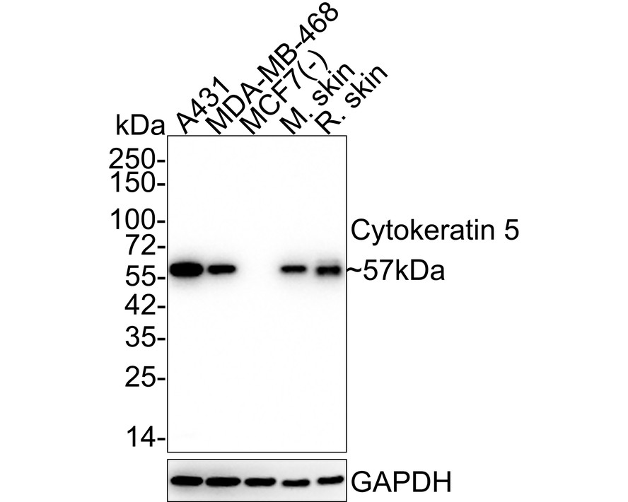
WB
Western blot analysis of Cytokeratin 5 on different lysates with Rabbit anti-Cytokeratin 5 antibody at 1/5,000 dilution. Lane 1: A431 cell lysate, Lane 2: MDA-MB-468 cell lysate, Lane 3: MCF7 cell lysate (negative), Lane 4: Mouse skin tissue lysate, Lane 5: Rat skin tissue lysate, Lysates/proteins at 20 µg/Lane. Exposure time: 2 minutes 6 seconds; 4-20% SDS-PAGE gel. Proteins were transferred to a PVDF membrane and blocked with 5% NFDM/TBST for 1 hour at room temperature. The primary antibody at 1/5,000 dilution was used in 5% NFDM/TBST at 4℃ overnight. Goat Anti-Rabbit IgG - HRP Secondary Antibody at 1/50,000 dilution was used for 1 hour at room temperature.IHC
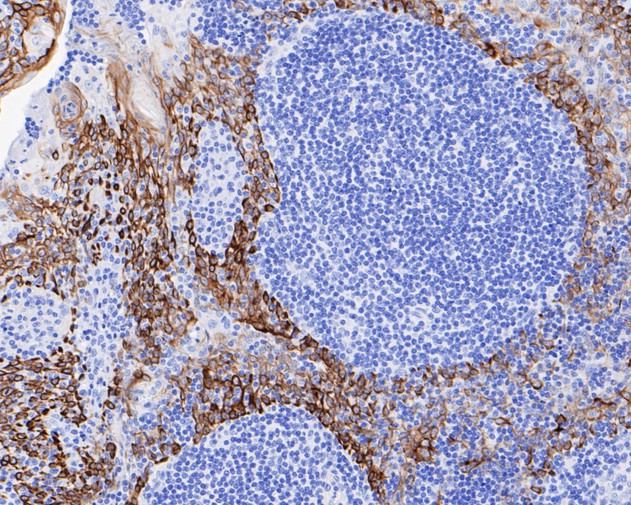
Immunohistochemical analysis of paraffin-embedded human tonsil tissue using anti-Cytokeratin 5 antibody. The section was pre-treated using heat mediated antigen retrieval with Tris-EDTA buffer (pH 9.0) for 20 minutes.The tissues were blocked in 5% BSA for 30 minutes at room temperature, washed with ddH2O and PBS, and then probed with the primary antibody (1/50) for 30 minutes at room temperature. The detection was performed using an HRP conjugated compact polymer system. DAB was used as the chromogen. Tissues were counterstained with hematoxylin and mounted with DPX.ICC/IF
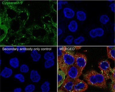
Immunocytochemistry analysis of A431 cells labeling Cytokeratin 5 with Rabbit anti-Cytokeratin 5 antibody at 1/500 dilution. Cells were fixed in 4% paraformaldehyde for 20 minutes at room temperature, permeabilized with 0.1% Triton X-100 in PBS for 5 minutes at room temperature, then blocked with 1% BSA in 10% negative goat serum for 1 hour at room temperature. Cells were then incubated with Rabbit anti-Cytokeratin 5 antibody at 1/500 dilution in 1% BSA in PBST overnight at 4 ℃. Goat Anti-Rabbit IgG H&L (iFluor™ 488) was used as the secondary antibody at 1/1,000 dilution. PBS instead of the primary antibody was used as the secondary antibody only control. Nuclear DNA was labelled in blue with DAPI. Beta tubulin (red) was stained at 1/100 dilution overnight at +4℃. Goat Anti-Mouse IgG H&L (iFluor™ 594,) was used as the secondary antibody at 1/1,000 dilution.IF-P
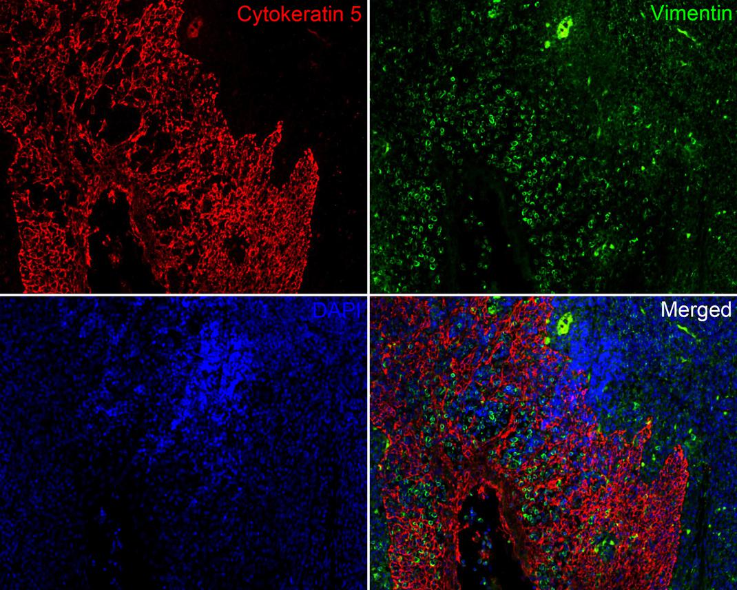
Immunofluorescence analysis of paraffin-embedded human tonsil tissue labeling Cytokeratin 5 at 1/200 dilution and Vimentin at 1/200 dilution. The section was pre-treated using heat mediated antigen retrieval with Tris-EDTA buffer (pH 9.0) for 20 minutes. The tissues were blocked in 10% negative goat serum for 1 hour at room temperature, washed with PBS. And then probed with the primary antibodies Cytokeratin 5 (red) at 1/200 dilution and Vimentin (green) at 1/200 dilution at +4℃ overnight, washed with PBS. iFluorTM 594 conjugate-Goat anti-Rabbit IgG and iFluorTM 488 conjugate-Goat anti-Mouse IgG were used as the secondary antibodies at 1/1000 dilution. DAPI was used as nuclear counterstain.FC
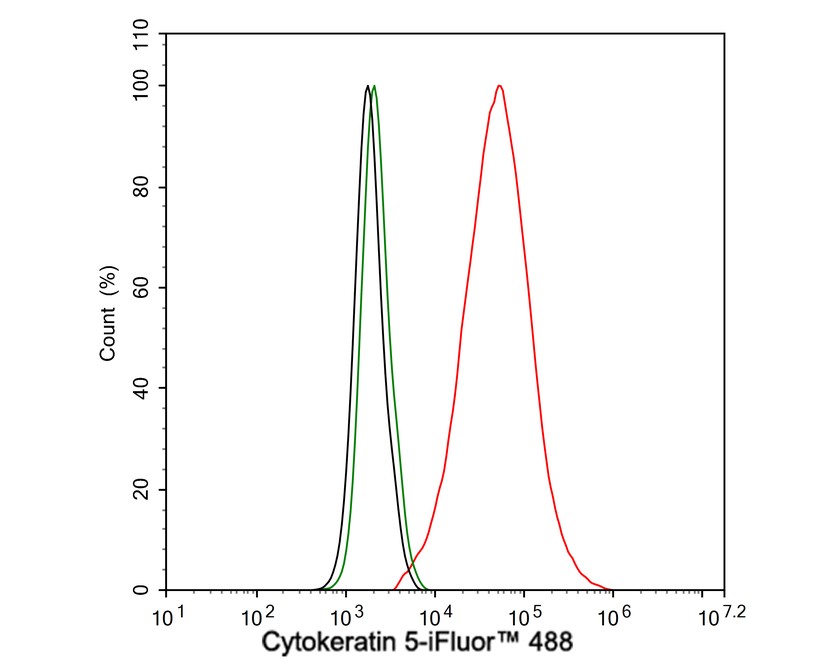
Flow cytometric analysis of A431 cells labeling Cytokeratin 5. Cells were fixed and permeabilized. Then stained with the primary antibody (1μg/mL) (red) compared with Rabbit IgG Isotype Control (green). After incubation of the primary antibody at +4℃ for an hour, the cells were stained with a iFluor™ 488 conjugate-Goat anti-Rabbit IgG Secondary antibody at 1/1,000 dilution for 30 minutes at +4℃. Unlabelled sample was used as a control (cells without incubation with primary antibody; black).| Product Name | Cytokeratin 5 Recombinant Rabbit Monoclonal Antibody |
|---|---|
| Antibody Type | Primary Antibodies |
| Product description | Cytokeratins comprise a diverse group of intermediate filament proteins (IFPs) that are expressed as pairs in both keratinized and non-keratinized epithelial tissue. Cytokeratins play a critical role in differentiation and tissue specialization and function to maintain the overall structural integrity of epithelial cells. Cytokeratins have been found to be useful markers of tissue differentiation which is directly applicable to the characterization of malignant tumors. Cytokeratin 5 is expressed in normal basal cells. Mutations of the cytokeratin 5 gene (KRT5) have been shown to result in the autosomal dominant disorder epidermolysis bullosa (EB). |
| Immunogen | recombinant protein |
| Clonality | monoclonal |
|---|---|
| Isotype | IgG |
| Host Species | Recombinant rabbit |
| Tested Applications | FCICC/IFIF-PIHCWB |
| WB:1:5000 ICC:1:500 IHC:1:50-1:500 IF-P:1:200 FC:1:1000 |
|
| Species Reactivity | HumanMouseRat |
| Concentration | 1mg/mL. |
| Purification | Protein A |
| Gene Symbol | KRT5 |
|---|---|
| Gene Synonyms | K5 CK5 DDD DDD1 EBS1 EBS2 EBS2A EBS2B EBS2C EBS2D EBS2E EBS2F KRT5A |
| Gene Full Name | keratin 5 |
| Alternative Names | 58 kDa cytokeratin antibody
CK-5 antibody
CK5 antibody
Cytokeratin-5 antibody
Cytokeratin5 antibody
DDD antibody
DDD1 antibody
EBS2 antibody
epidermolysis bullosa simplex 2 Dowling-Meara/Kobner/Weber-Cockayne types antibody
K2C5_HUMAN antibody
K5 antibody
keratin 5 (epidermolysis bullosa simplex Dowling-Meara/Kobner/Weber-Cockayne types) antibody Keratin 5 antibody Keratin antibody keratin complex 2 basic gene 5 antibody keratin type II cytoskeletal 5 antibody Keratin-5 antibody Keratin5 antibody KRT 5 antibody Krt5 antibody KRT5A antibody type II cytoskeletal 5 antibody Type-II keratin Kb5 antibody |
| Molecular Weight(MW) | 62kDa(Observed band size: 57kDa) |
| Cellular Localization | Cytoplasm. |
| SwissProt ID | P13647 |
|---|
WB
Western blot analysis of Cytokeratin 5 on different lysates with Rabbit anti-Cytokeratin 5 antibody at 1/5,000 dilution. Lane 1: A431 cell lysate, Lane 2: MDA-MB-468 cell lysate, Lane 3: MCF7 cell lysate (negative), Lane 4: Mouse skin tissue lysate, Lane 5: Rat skin tissue lysate, Lysates/proteins at 20 µg/Lane. Exposure time: 2 minutes 6 seconds; 4-20% SDS-PAGE gel. Proteins were transferred to a PVDF membrane and blocked with 5% NFDM/TBST for 1 hour at room temperature. The primary antibody at 1/5,000 dilution was used in 5% NFDM/TBST at 4℃ overnight. Goat Anti-Rabbit IgG - HRP Secondary Antibody at 1/50,000 dilution was used for 1 hour at room temperature.
IHC
Immunohistochemical analysis of paraffin-embedded human tonsil tissue using anti-Cytokeratin 5 antibody. The section was pre-treated using heat mediated antigen retrieval with Tris-EDTA buffer (pH 9.0) for 20 minutes.The tissues were blocked in 5% BSA for 30 minutes at room temperature, washed with ddH2O and PBS, and then probed with the primary antibody (1/50) for 30 minutes at room temperature. The detection was performed using an HRP conjugated compact polymer system. DAB was used as the chromogen. Tissues were counterstained with hematoxylin and mounted with DPX.
ICC/IF
Immunocytochemistry analysis of A431 cells labeling Cytokeratin 5 with Rabbit anti-Cytokeratin 5 antibody at 1/500 dilution. Cells were fixed in 4% paraformaldehyde for 20 minutes at room temperature, permeabilized with 0.1% Triton X-100 in PBS for 5 minutes at room temperature, then blocked with 1% BSA in 10% negative goat serum for 1 hour at room temperature. Cells were then incubated with Rabbit anti-Cytokeratin 5 antibody at 1/500 dilution in 1% BSA in PBST overnight at 4 ℃. Goat Anti-Rabbit IgG H&L (iFluor™ 488) was used as the secondary antibody at 1/1,000 dilution. PBS instead of the primary antibody was used as the secondary antibody only control. Nuclear DNA was labelled in blue with DAPI. Beta tubulin (red) was stained at 1/100 dilution overnight at +4℃. Goat Anti-Mouse IgG H&L (iFluor™ 594,) was used as the secondary antibody at 1/1,000 dilution.
IF-P
Immunofluorescence analysis of paraffin-embedded human tonsil tissue labeling Cytokeratin 5 at 1/200 dilution and Vimentin at 1/200 dilution. The section was pre-treated using heat mediated antigen retrieval with Tris-EDTA buffer (pH 9.0) for 20 minutes. The tissues were blocked in 10% negative goat serum for 1 hour at room temperature, washed with PBS. And then probed with the primary antibodies Cytokeratin 5 (red) at 1/200 dilution and Vimentin (green) at 1/200 dilution at +4℃ overnight, washed with PBS. iFluorTM 594 conjugate-Goat anti-Rabbit IgG and iFluorTM 488 conjugate-Goat anti-Mouse IgG were used as the secondary antibodies at 1/1000 dilution. DAPI was used as nuclear counterstain.
FC
Flow cytometric analysis of A431 cells labeling Cytokeratin 5. Cells were fixed and permeabilized. Then stained with the primary antibody (1μg/mL) (red) compared with Rabbit IgG Isotype Control (green). After incubation of the primary antibody at +4℃ for an hour, the cells were stained with a iFluor™ 488 conjugate-Goat anti-Rabbit IgG Secondary antibody at 1/1,000 dilution for 30 minutes at +4℃. Unlabelled sample was used as a control (cells without incubation with primary antibody; black).| Positive Control | A431, human tonsil tissue, human liver cancer tissue, human breast carcinoma tissue. |
|---|---|
| Application Notes | WB:1:5000 ICC:1:500 IHC:1:50-1:500 IF-P:1:200 FC:1:1000 |
| Form | Liquid |
|---|---|
| Storage Instructions | Store at +4℃ after thawing. Aliquot store at -20℃ or -80℃. Avoid repeated freeze / thaw cycles. |
| Storage Buffer | 1*TBS (pH7.4), 1%BSA, 40%Glycerol. Preservative: 0.05% Sodium Azide. |


 Data sheet for OM637368