WB
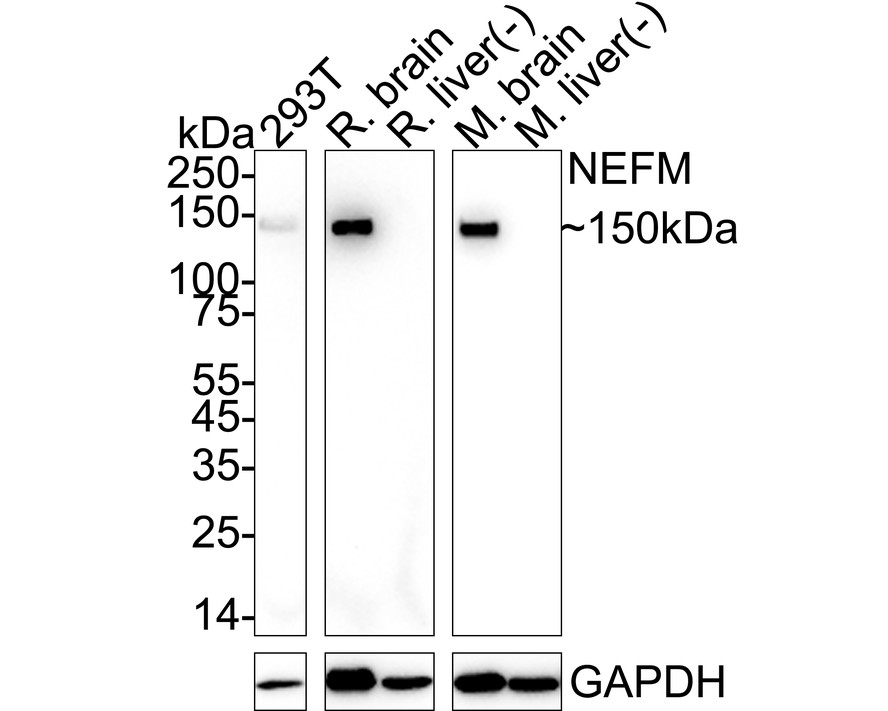
Western blot analysis of NF-M on different lysates with Rabbit anti-NF-M antibody at 1/1,000 dilution. Lane 1: 293T cell lysate (20 µg/Lane), Lane 2: Rat brain tissue lysate (40 µg/Lane), Lane 3: Rat liver tissue lysate (negative) (40 µg/Lane), Lane 4: Mouse brain tissue lysate (40 µg/Lane), Lane 5: Mouse liver tissue lysate (negative) (40 µg/Lane), Exposure time: Lane 1-3: 3 minutes; Lane 4-5: 18 seconds; 4-20% SDS-PAGE gel. Proteins were transferred to a PVDF membrane and blocked with 5% NFDM/TBST for 1 hour at room temperature. The primary antibody at 1/1,000 dilution was used in 5% NFDM/TBST at 4℃ overnight. Goat Anti-Rabbit IgG - HRP Secondary Antibody at 1/50,000 dilution was used for 1 hour at room temperature.IHC
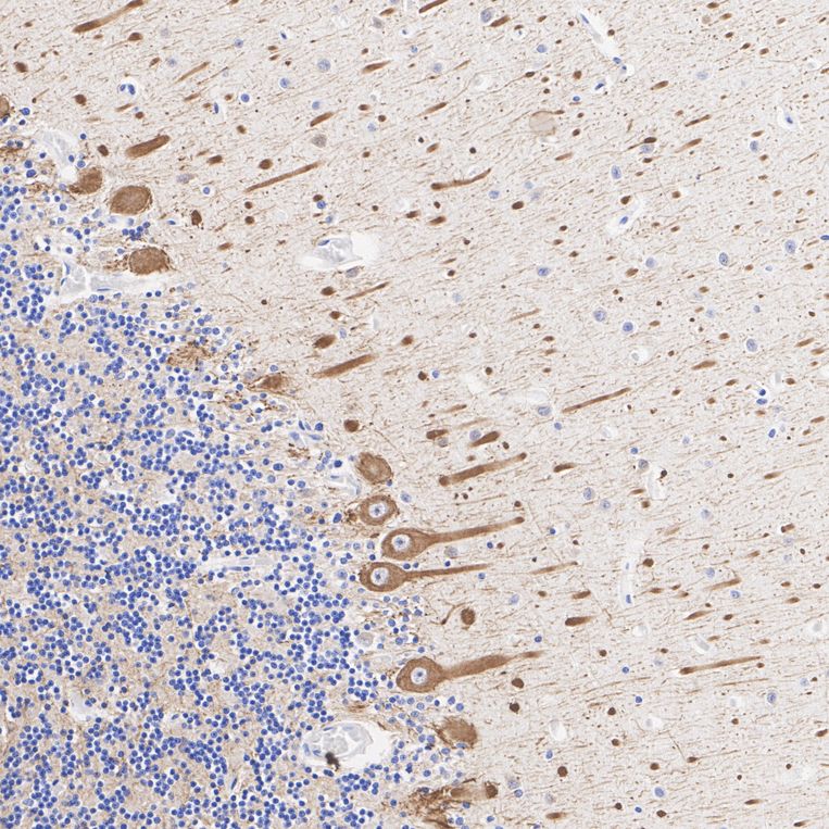
Immunohistochemical analysis of paraffin-embedded human cerebellum tissue with Rabbit anti-NF-M antibody at 1/2,000 dilution. The section was pre-treated using heat mediated antigen retrieval with Tris-EDTA buffer (pH 9.0) for 20 minutes. The tissues were blocked in 1% BSA for 20 minutes at room temperature, washed with ddH2O and PBS, and then probed with the primary antibody at 1/2,000 dilution for 1 hour at room temperature. The detection was performed using an HRP conjugated compact polymer system. DAB was used as the chromogen. Tissues were counterstained with hematoxylin and mounted with DPX.IHC
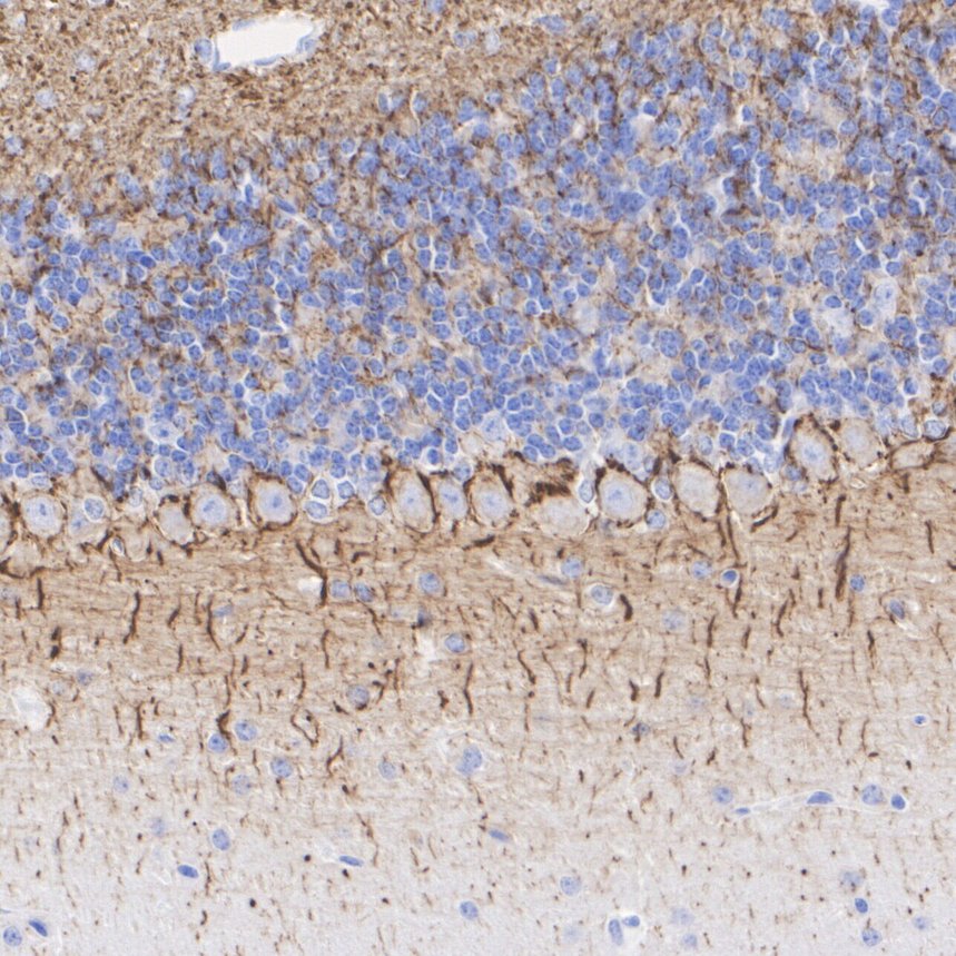
Immunohistochemical analysis of paraffin-embedded mouse cerebellum tissue with Rabbit anti-NF-M antibody at 1/2,000 dilution. The section was pre-treated using heat mediated antigen retrieval with Tris-EDTA buffer (pH 9.0) for 20 minutes. The tissues were blocked in 1% BSA for 20 minutes at room temperature, washed with ddH2O and PBS, and then probed with the primary antibody at 1/2,000 dilution for 1 hour at room temperature. The detection was performed using an HRP conjugated compact polymer system. DAB was used as the chromogen. Tissues were counterstained with hematoxylin and mounted with DPX.IHC
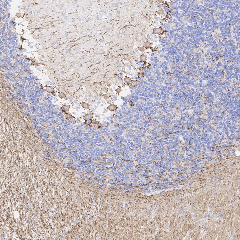
Immunohistochemical analysis of paraffin-embedded rat cerebellum tissue with Rabbit anti-NF-M antibody at 1/2,000 dilution. The section was pre-treated using heat mediated antigen retrieval with Tris-EDTA buffer (pH 9.0) for 20 minutes. The tissues were blocked in 1% BSA for 20 minutes at room temperature, washed with ddH2O and PBS, and then probed with the primary antibody at 1/2,000 dilution for 1 hour at room temperature. The detection was performed using an HRP conjugated compact polymer system. DAB was used as the chromogen. Tissues were counterstained with hematoxylin and mounted with DPX.IF-F
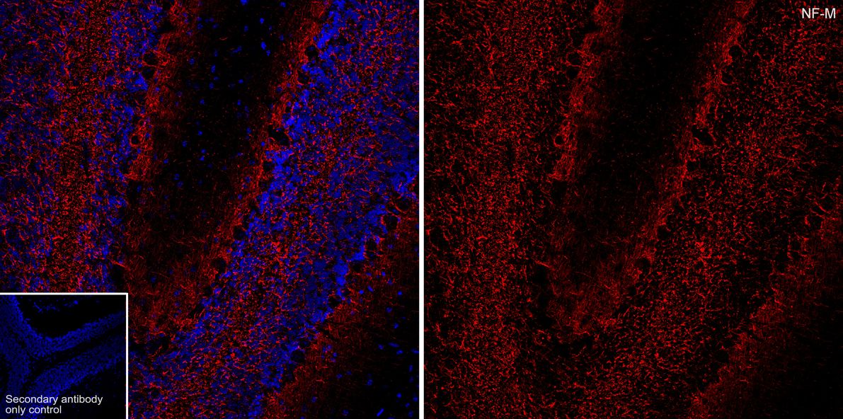
Species: Mouse, Site: Cerebellum, Sample: Frozen section, Antibody concentration: 1:500, Antigen retrieval: Not required.IF-F
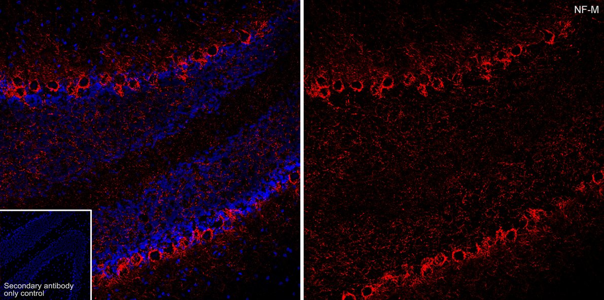
Species: Rat, Site: Cerebellum, Sample: Frozen section, Antibody concentration: 1:500, Antigen retrieval: Not required.| Product Name | NF-M Recombinant Rabbit Monoclonal Antibody |
|---|---|
| Antibody Type | Primary Antibodies |
| Product description | Neurofilament-M (NF-M), for neurofilament medium polypeptide, a member of the intermediate filament family, is a major component of neuronal cytoskeletons. Neurofilaments are dynamic structures; they contain phosphorylation sites for a large number of protein kinases, including protein kinase A, protein kinase C, cyclin-dependent kinase 5, extracellular signal regulated kinase, glycogen synthase kinase-3, and stress-activated protein kinase gamma. In addition to their role in the control of axon caliber, neurofilaments may affect other cytoskeletal elements, such as microtubules and actin filaments. Changes in neurofilament phosphorylation or metabolism are frequently observed in neurodegenerative diseases, including amyotrophic lateral sclerosis (ALS), Parkinson's disease, and Alzheimer's disease. |
| Immunogen | Synthetic peptide within Human NEFM aa 651-692 / 916. |
| Clonality | Monoclonal |
|---|---|
| Isotype | IgG |
| Host Species | Recombinant rabbit |
| Tested Applications | IF-FIHCWB |
| WB:1:1000-1:2000 IHC:1:2000 IF-F:1:500 |
|
| Species Reactivity | HumanMouseRat |
| Concentration | 1mg/ml |
| Purification | Protein A |
| Gene Symbol | NEFM |
|---|---|
| Gene Synonyms | NFM NEF3 NF-M |
| Gene Full Name | neurofilament medium chain |
| Gene Summary | Neurofilaments are type IV intermediate filament heteropolymers composed of light, medium, and heavy chains. Neurofilaments comprise the axoskeleton and functionally maintain neuronal caliber. They may also play a role in intracellular transport to axons and dendrites. This gene encodes the medium neurofilament protein. This protein is commonly used as a biomarker of neuronal damage. Alternative splicing results in multiple transcript variants encoding distinct isoforms. [provided by RefSeq, Oct 2008] |
| Molecular Weight(MW) | 102kDa(Observed band size: 150kDa) |
| Cellular Localization | Cell projection, Cytoplasm, Cytoskeleton, Intermediate filament. |
WB
Western blot analysis of NF-M on different lysates with Rabbit anti-NF-M antibody at 1/1,000 dilution. Lane 1: 293T cell lysate (20 µg/Lane), Lane 2: Rat brain tissue lysate (40 µg/Lane), Lane 3: Rat liver tissue lysate (negative) (40 µg/Lane), Lane 4: Mouse brain tissue lysate (40 µg/Lane), Lane 5: Mouse liver tissue lysate (negative) (40 µg/Lane), Exposure time: Lane 1-3: 3 minutes; Lane 4-5: 18 seconds; 4-20% SDS-PAGE gel. Proteins were transferred to a PVDF membrane and blocked with 5% NFDM/TBST for 1 hour at room temperature. The primary antibody at 1/1,000 dilution was used in 5% NFDM/TBST at 4℃ overnight. Goat Anti-Rabbit IgG - HRP Secondary Antibody at 1/50,000 dilution was used for 1 hour at room temperature.
IHC
Immunohistochemical analysis of paraffin-embedded human cerebellum tissue with Rabbit anti-NF-M antibody at 1/2,000 dilution. The section was pre-treated using heat mediated antigen retrieval with Tris-EDTA buffer (pH 9.0) for 20 minutes. The tissues were blocked in 1% BSA for 20 minutes at room temperature, washed with ddH2O and PBS, and then probed with the primary antibody at 1/2,000 dilution for 1 hour at room temperature. The detection was performed using an HRP conjugated compact polymer system. DAB was used as the chromogen. Tissues were counterstained with hematoxylin and mounted with DPX.
IHC
Immunohistochemical analysis of paraffin-embedded mouse cerebellum tissue with Rabbit anti-NF-M antibody at 1/2,000 dilution. The section was pre-treated using heat mediated antigen retrieval with Tris-EDTA buffer (pH 9.0) for 20 minutes. The tissues were blocked in 1% BSA for 20 minutes at room temperature, washed with ddH2O and PBS, and then probed with the primary antibody at 1/2,000 dilution for 1 hour at room temperature. The detection was performed using an HRP conjugated compact polymer system. DAB was used as the chromogen. Tissues were counterstained with hematoxylin and mounted with DPX.
IHC
Immunohistochemical analysis of paraffin-embedded rat cerebellum tissue with Rabbit anti-NF-M antibody at 1/2,000 dilution. The section was pre-treated using heat mediated antigen retrieval with Tris-EDTA buffer (pH 9.0) for 20 minutes. The tissues were blocked in 1% BSA for 20 minutes at room temperature, washed with ddH2O and PBS, and then probed with the primary antibody at 1/2,000 dilution for 1 hour at room temperature. The detection was performed using an HRP conjugated compact polymer system. DAB was used as the chromogen. Tissues were counterstained with hematoxylin and mounted with DPX.
IF-F
Species: Mouse, Site: Cerebellum, Sample: Frozen section, Antibody concentration: 1:500, Antigen retrieval: Not required.
IF-F
Species: Rat, Site: Cerebellum, Sample: Frozen section, Antibody concentration: 1:500, Antigen retrieval: Not required.| Application Notes | WB:1:1000-1:2000 IHC:1:2000 IF-F:1:500 |
|---|
| Form | Liquid |
|---|---|
| Storage Instructions | Store at +4℃ after thawing. Aliquot store at -20℃ or -80℃. Avoid repeated freeze / thaw cycles. |
| Storage Buffer | 1*TBS (pH7.4), 1%BSA, 40%Glycerol. Preservative: 0.05% Sodium Azide. |


