Application
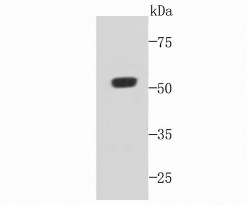
Fig1: Western blot analysis of C19orf35 on recombinant protein using anti-C19orf35 antibody at 1/1,000 dilution.Application
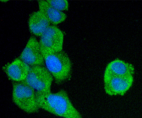
Fig2: ICC staining C19orf35 in Hela cells (green). The nuclear counter stain is DAPI (blue). Cells were fixed in paraformaldehyde, permeabilised with 0.25% Triton X100/PBS.Application
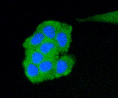
Fig3: ICC staining C19orf35 in MCF-7 cells (green). The nuclear counter stain is DAPI (blue). Cells were fixed in paraformaldehyde, permeabilised with 0.25% Triton X100/PBS.Application
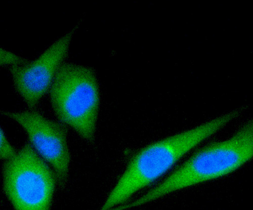
Fig4: ICC staining C19orf35 in PC-3M cells (green). The nuclear counter stain is DAPI (blue). Cells were fixed in paraformaldehyde, permeabilised with 0.25% Triton X100/PBS.Application
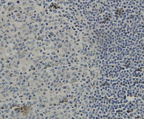
Fig5: Immunohistochemical analysis of paraffin-embedded human tonsil tissue using anti-C19orf35 antibody. Counter stained with hematoxylin.Application
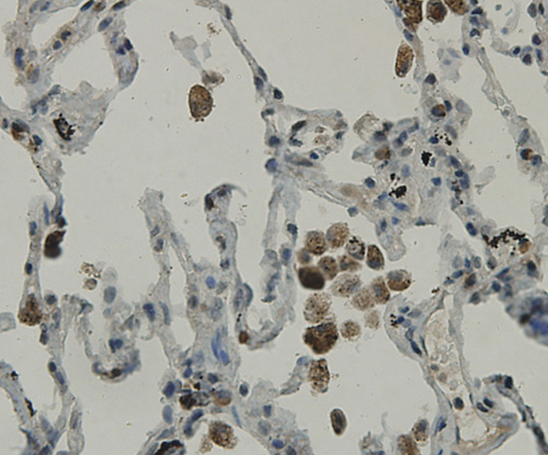
Fig6: Immunohistochemical analysis of paraffin-embedded human lung tissue using anti-C19orf35 antibody. Counter stained with hematoxylin.Application
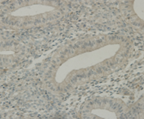
Fig7: Immunohistochemical analysis of paraffin-embedded human uterus tissue using anti-C19orf35 antibody. Counter stained with hematoxylin.Application
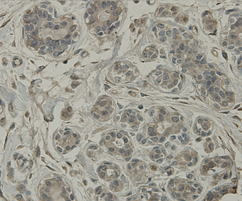
Fig8: Immunohistochemical analysis of paraffin-embedded human breast tissue using anti-C19orf35 antibody. Counter stained with hematoxylin.| Product Name | C19orf35 [3-B7-G3] |
|---|---|
| Antibody Type | Primary Antibodies |
| Antigen Alias | C19orf35 antibody Chromosome 19 open reading frame 35 antibody CS035_HUMAN antibody FLJ45778 antibody Putative uncharacterized protein C19orf35 antibody |
| Product description | C19orf35 and CNRIP1 were specific to diffuse-type compared to the intestinal-type subgroups. Both revealed a similar level of methylation in CNM regardless of the histologic subtype of tumor while the methylation of the tumor tissue varied with the histologic type. C19orf35 was hypermethylated but CNRIP1 was hypomethylated in diffuse-type tumors compared to the CNM while it had an opposite trend in the tumors of the intestinal-type and mixed-type subgroups. Importantly, research suggest the possibility of utilizing these genes as specific markers for diffuse-type GCs. On the other hand, the function of C19orf35 is still unknown while frequent methylation of CNRIP1 has recently been reported in colorectal cancers and adenoma. |
| Immunogen | Recombinant protein |
| Clonality | Monoclonal |
|---|---|
| Isotype | IgG1 |
| Host Species | Mouse |
| Tested Applications | WBICCIHC |
| WB:1:250~1:1000 ICC:1:100~1:200 IHC:1:20~1:100 Notes:Optimal dilutions/concentrations should be determined by the researcher. |
|
| Species Reactivity | Human |
| Concentration | 2mg/ml |
| Alternative Names | C19orf35 antibody Chromosome 19 open reading frame 35 antibody CS035_HUMAN antibody FLJ45778 antibody Putative uncharacterized protein C19orf35 antibody |
|---|---|
| Molecular Weight(MW) | 51 kDa |
| Cellular Localization | Actin cytoskeleton. |
| SwissProt ID | Q6ZS72 |
|---|
Application
Fig1: Western blot analysis of C19orf35 on recombinant protein using anti-C19orf35 antibody at 1/1,000 dilution.
Application
Fig2: ICC staining C19orf35 in Hela cells (green). The nuclear counter stain is DAPI (blue). Cells were fixed in paraformaldehyde, permeabilised with 0.25% Triton X100/PBS.
Application
Fig3: ICC staining C19orf35 in MCF-7 cells (green). The nuclear counter stain is DAPI (blue). Cells were fixed in paraformaldehyde, permeabilised with 0.25% Triton X100/PBS.
Application
Fig4: ICC staining C19orf35 in PC-3M cells (green). The nuclear counter stain is DAPI (blue). Cells were fixed in paraformaldehyde, permeabilised with 0.25% Triton X100/PBS.
Application
Fig5: Immunohistochemical analysis of paraffin-embedded human tonsil tissue using anti-C19orf35 antibody. Counter stained with hematoxylin.
Application
Fig6: Immunohistochemical analysis of paraffin-embedded human lung tissue using anti-C19orf35 antibody. Counter stained with hematoxylin.
Application
Fig7: Immunohistochemical analysis of paraffin-embedded human uterus tissue using anti-C19orf35 antibody. Counter stained with hematoxylin.
Application
Fig8: Immunohistochemical analysis of paraffin-embedded human breast tissue using anti-C19orf35 antibody. Counter stained with hematoxylin.| Positive Control | Recombinant protein, Hela, MCF-7, PC-3M, human tonsil tissue, human lung tissue, human uterus tissue, human breast tissue. |
|---|---|
| Application Notes | WB:1:250~1:1000 ICC:1:100~1:200 IHC:1:20~1:100 Notes:Optimal dilutions/concentrations should be determined by the researcher. |
| Form | Liquid |
|---|---|
| Storage Instructions | Store at +4℃ after thawing. Aliquot store at -20℃ or -80℃. Avoid repeated freeze / thaw cycles. |
| Storage Buffer | 1*TBS (pH7.4), 1%BSA, 40%Glycerol. Preservative: 0.05% Sodium Azide. |


