Application
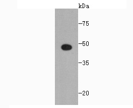
Fig1: Western blot analysis of SCAI on Recombinant protein using anti-SCAI antibody at 1/1,000 dilution.Application
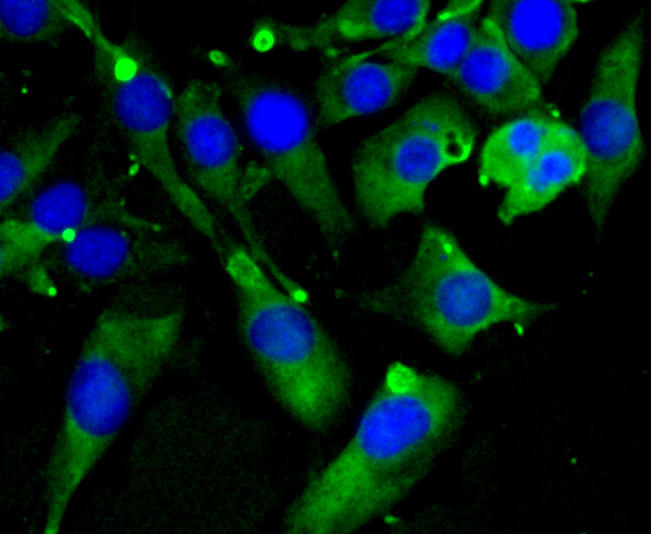
Fig2: ICC staining SCAI in SHG-44 cells (green). The nuclear counter stain is DAPI (blue). Cells were fixed in paraformaldehyde, permeabilised with 0.25% Triton X100/PBS.Application
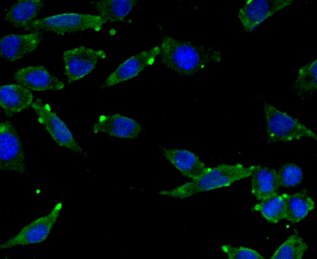
Fig3: ICC staining SCAI in SH-SY-5Y cells (green). The nuclear counter stain is DAPI (blue). Cells were fixed in paraformaldehyde, permeabilised with 0.25% Triton X100/PBS.Application
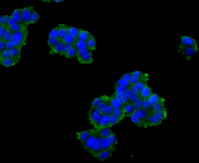
Fig4: ICC staining SCAI in PC-12 cells (green). The nuclear counter stain is DAPI (blue). Cells were fixed in paraformaldehyde, permeabilised with 0.25% Triton X100/PBS.Application
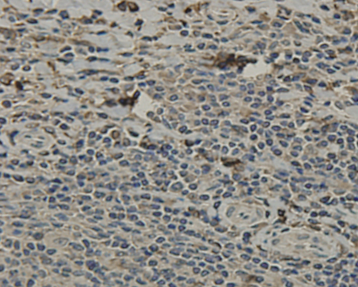
Fig5: Immunohistochemical analysis of paraffin-embedded human tonsil tissue using anti-SCAI antibody. Counter stained with hematoxylin.Application
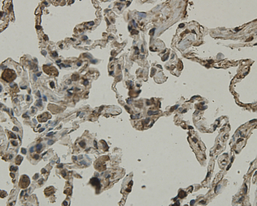
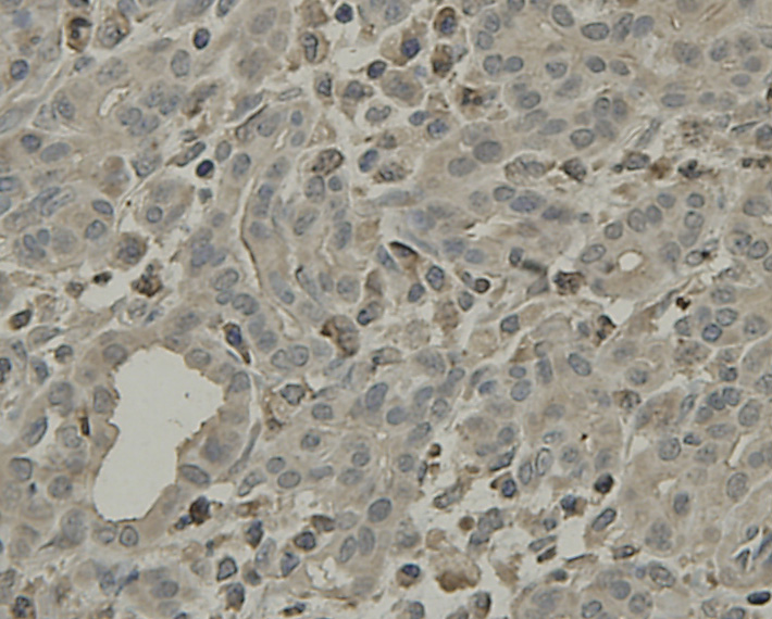
Fig6: Immunohistochemical analysis of paraffin-embedded human lung tissue using anti-SCAI antibody. Counter stained with hematoxylin.Application
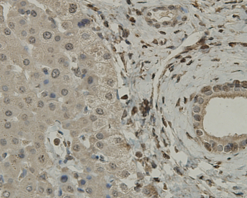
Fig7: Immunohistochemical analysis of paraffin-embedded human liver tissue using anti-SCAI antibody. Counter stained with hematoxylin.Application
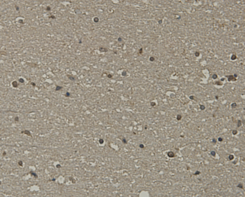
Fig8: Immunohistochemical analysis of paraffin-embedded human brain tissue using anti-SCAI antibody. Counter stained with hematoxylin.Application
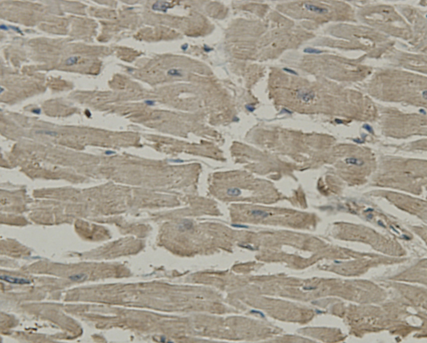
Fig9: Immunohistochemical analysis of paraffin-embedded human heart tissue using anti-SCAI antibody. Counter stained with hematoxylin.Application
Fig10: Immunohistochemical analysis of paraffin-embedded human pancreas tissue using anti-SCAI antibody. Counter stained with hematoxylin.| Product Name | SCAI [7-A4] |
|---|---|
| Antibody Type | Primary Antibodies |
| Antigen Alias | C9orf126 antibody FLJ11758 antibody FLJ36664 antibody MGC120525 antibody MGC120526 antibody MGC120528 antibody NET40 antibody Protein SCAI antibody SCAI antibody SCAI_HUMAN antibody Suppressor of cancer cell invasion antibody Suppressor of cancer cell invasion protein antibody |
| Product description | Protein SCAI, also known as Suppressor of cancer cell invasion protein, and encoded by the gene name SCAI, is a tumor suppressor that suppresses MKL1-induced SRF transcriptional activity. It is a regulator of cell migration and appears to be involved in the RhoA (ras homolog gene family, member A)-Dia1 (diaphanous homolog 1) signal transduction pathway. SCAI localizes in the nucleus, where it binds and inhibits the myocardin-related transcription factor MAL by forming a ternary complex with serum response factor (SRF). |
| Immunogen | Recombinant protein |
| Clonality | Monoclonal |
|---|---|
| Isotype | IgM |
| Host Species | Mouse |
| Tested Applications | WBICCIHC |
| WB:1:1,000-1:2,000 ICC:1:50-1:200 IHC:1:50-1:200 |
|
| Species Reactivity | Human |
| Concentration | 2mg/ml |
| Alternative Names | C9orf126 antibody FLJ11758 antibody FLJ36664 antibody MGC120525 antibody MGC120526 antibody MGC120528 antibody NET40 antibody Protein SCAI antibody SCAI antibody SCAI_HUMAN antibody Suppressor of cancer cell invasion antibody Suppressor of cancer cell invasion protein antibody |
|---|---|
| Molecular Weight(MW) | 48 kDa |
| Cellular Localization | Membrane. Nucleus. Cytoplasm. |
| SwissProt ID | Q8N9R8 |
|---|
Application
Fig1: Western blot analysis of SCAI on Recombinant protein using anti-SCAI antibody at 1/1,000 dilution.
Application
Fig2: ICC staining SCAI in SHG-44 cells (green). The nuclear counter stain is DAPI (blue). Cells were fixed in paraformaldehyde, permeabilised with 0.25% Triton X100/PBS.
Application
Fig3: ICC staining SCAI in SH-SY-5Y cells (green). The nuclear counter stain is DAPI (blue). Cells were fixed in paraformaldehyde, permeabilised with 0.25% Triton X100/PBS.
Application
Fig4: ICC staining SCAI in PC-12 cells (green). The nuclear counter stain is DAPI (blue). Cells were fixed in paraformaldehyde, permeabilised with 0.25% Triton X100/PBS.
Application
Fig5: Immunohistochemical analysis of paraffin-embedded human tonsil tissue using anti-SCAI antibody. Counter stained with hematoxylin.
Application
Fig6: Immunohistochemical analysis of paraffin-embedded human lung tissue using anti-SCAI antibody. Counter stained with hematoxylin.
Application
Fig7: Immunohistochemical analysis of paraffin-embedded human liver tissue using anti-SCAI antibody. Counter stained with hematoxylin.
Application
Fig8: Immunohistochemical analysis of paraffin-embedded human brain tissue using anti-SCAI antibody. Counter stained with hematoxylin.
Application
Fig9: Immunohistochemical analysis of paraffin-embedded human heart tissue using anti-SCAI antibody. Counter stained with hematoxylin.
Application
Fig10: Immunohistochemical analysis of paraffin-embedded human pancreas tissue using anti-SCAI antibody. Counter stained with hematoxylin.| Positive Control | Recombinant protein, SHG-44, SH-SY-5Y, PC-12, human tonsil tissue, human lung tissue, human liver tissue, human brain tissue, human heart tissue, human pancreas tissue. |
|---|---|
| Application Notes | WB:1:1,000-1:2,000 ICC:1:50-1:200 IHC:1:50-1:200 |
| Form | Liquid |
|---|---|
| Storage Instructions | Store at -20 ℃. Stable for 12 months from date of receipt. |
| Storage Buffer | 1*TBS (pH7.4), 1%BSA, 40%Glycerol. Preservative: 0.05% Sodium Azide. |


