Application
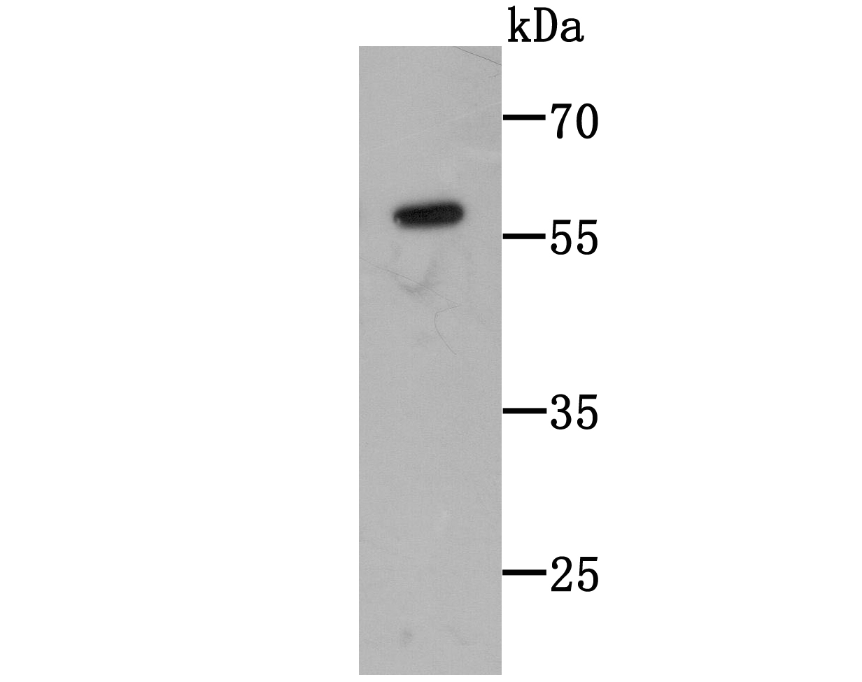
Fig1: Western blot analysis of C14orf93 on C14orf93-GST recombinant protein lysate using anti-C14orf93 antibody at 1/1,000 dilution.Application
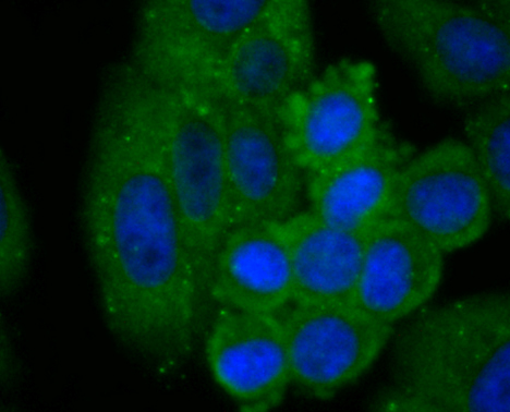
Fig2: ICC staining C14orf93 in HepG2 cells (green). The nuclear counter stain is DAPI (blue). Cells were fixed in paraformaldehyde, permeabilised with 0.25% Triton X100/PBS.Application
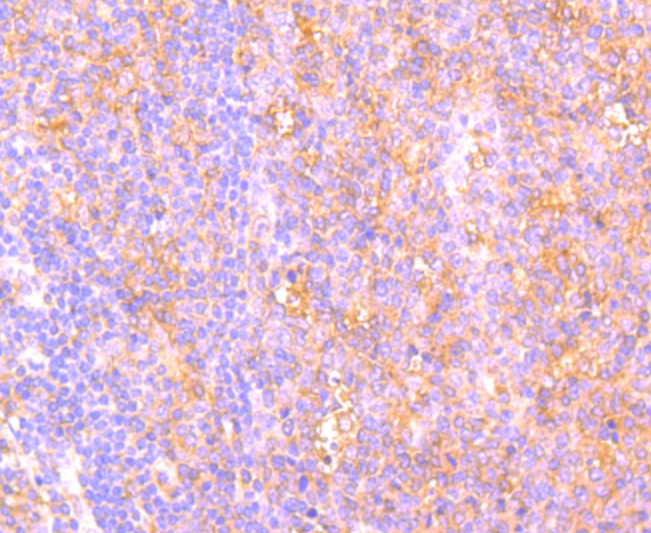
Fig3: Immunohistochemical analysis of paraffin-embedded human spleen tissue using anti-C14orf93 antibody. Counter stained with hematoxylin.Application
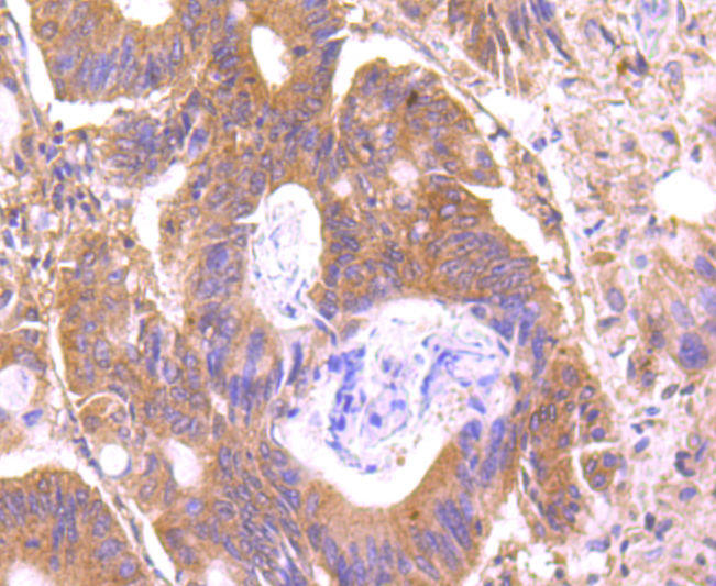
Fig4: Immunohistochemical analysis of paraffin-embedded human colon cancer tissue using anti-C14orf93 antibody. Counter stained with hematoxylin.Application
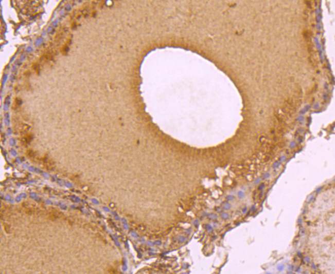
Fig5: Immunohistochemical analysis of paraffin-embedded human thyroid tissue using anti-C14orf93 antibody. Counter stained with hematoxylin.Application
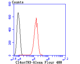
Fig6: Flow cytometric analysis of SH-SY5Y cells with C14orf93 antibody at 1/100 dilution (red) compared with an unlabelled control (cells without incubation with primary antibody; black).| Product Name | C14orf93 [2-F6] |
|---|---|
| Antibody Type | Primary Antibodies |
| Antigen Alias | C14orf93 antibody CN093_HUMAN antibody Uncharacterized protein C14orf93 antibody Uncharacterized protein C14orf93 homolog antibody |
| Product description | C14orf93 (also named as Regulator of Thyroid Function and Cancer, RTFC) as a novel susceptibility gene for familial nonmedullary thyroid cancer.The oncogenic functions of R115Q, V205M, and G209D RTFC mutants are demonstrated by cell surviving assay, migration assay, and colony forming assays. Moreover, RTFC has been identified as a potential antigen associated with the pathogenesis of peripheral T-cell lymphomas, not otherwise specified (PTCL, NOS). Two in vitro biochemical screens suggested that RTFC might have RNA and phosphopeptide (pSer/pThr-X-X-X-pSer/pThr) binding activities. Yet, the role of RTFC in normal development, as well as the molecular function of RTFC, remain unexplored. |
| Immunogen | Recombinant protein |
| Clonality | Monoclonal |
|---|---|
| Isotype | IgG1 |
| Host Species | Mouse |
| Tested Applications | WBICCIHCFC |
| WB:1:500-1:1,000 ICC:1:50-1:200 IHC:1:50-1:200 FC:1:50-1:200 |
|
| Species Reactivity | Human |
| Concentration | 2mg/ml |
| Alternative Names | C14orf93 antibody CN093_HUMAN antibody Uncharacterized protein C14orf93 antibody Uncharacterized protein C14orf93 homolog antibody |
|---|---|
| Molecular Weight(MW) | 57 kDa |
| Cellular Localization | Secreted. Extracellular region. |
| SwissProt ID | Q9H972 |
|---|
Application
Fig1: Western blot analysis of C14orf93 on C14orf93-GST recombinant protein lysate using anti-C14orf93 antibody at 1/1,000 dilution.
Application
Fig2: ICC staining C14orf93 in HepG2 cells (green). The nuclear counter stain is DAPI (blue). Cells were fixed in paraformaldehyde, permeabilised with 0.25% Triton X100/PBS.
Application
Fig3: Immunohistochemical analysis of paraffin-embedded human spleen tissue using anti-C14orf93 antibody. Counter stained with hematoxylin.
Application
Fig4: Immunohistochemical analysis of paraffin-embedded human colon cancer tissue using anti-C14orf93 antibody. Counter stained with hematoxylin.
Application
Fig5: Immunohistochemical analysis of paraffin-embedded human thyroid tissue using anti-C14orf93 antibody. Counter stained with hematoxylin.
Application
Fig6: Flow cytometric analysis of SH-SY5Y cells with C14orf93 antibody at 1/100 dilution (red) compared with an unlabelled control (cells without incubation with primary antibody; black).| Positive Control | Human placenta tissue lysate, HepG2, human spleen tissue, human colon cancer tissue, human thyroid tissue, SH-SY5Y cell. |
|---|---|
| Application Notes | WB:1:500-1:1,000 ICC:1:50-1:200 IHC:1:50-1:200 FC:1:50-1:200 |
| Form | Liquid |
|---|---|
| Storage Instructions | Store at +4℃after thawing. Aliquot store at -20℃or -80℃Avoid repeated freeze / thaw cycles. |
| Storage Buffer | 1*TBS (pH7.4), 0.5%BSA, 50%Glycerol. Preservative: 0.05% Sodium Azide. |


