Application
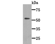
Fig1: Western blot analysis of WSCD2 on WSCD2 recombinant protein lysate using anti-WSCD2 antibody at 1/2000 dilution.Application
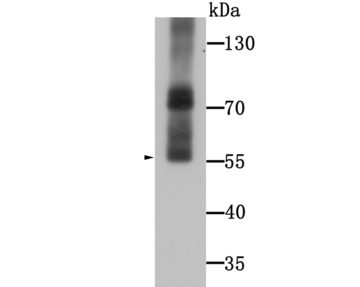
Fig2: Western blot analysis of WSCD2 on human stomach tissue lysate using anti-WSCD2 antibody at 1/1000 dilution.Application
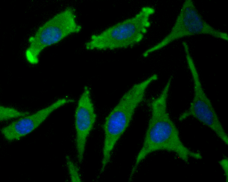
Fig3: ICC staining WSCD2 in SH-SY5Y cells (green). The nuclear counter stain is DAPI (blue). Cells were fixed in paraformaldehyde, permeabilised with 0.25% Triton X100/PBS.Application
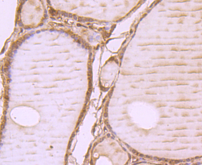
Fig4: Immunohistochemical analysis of paraffin-embedded human thyroid tissue using anti-WSCD2 antibody. Counter stained withApplication
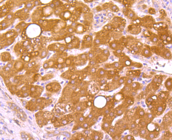
Fig5: Immunohistochemical analysis of paraffin-embedded human liver cancer tissue using anti-WSCD2 antibody. Counter stained with hematoxylin.Application
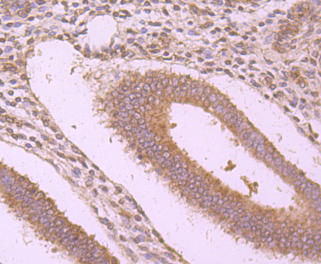
Fig6: Immunohistochemical analysis of paraffin-embedded human uterus tissue using anti-WSCD2 antibody. Counter stained with hematoxylin.| Product Name | WSCD2 [9-F3] |
|---|---|
| Antibody Type | Primary Antibodies |
| Antigen Alias | 4933413A10Rik antibody C530024P05Rik antibody Gm450 antibody WSC domain containing 2 antibody WSC domain containing protein 2 antibody WSC domain-containing protein 2 antibody WSCD 2 antibody WSCD2 antibody WSCD2_HUMAN antibody |
| Product description | WSC domain-containing protein 2 (WSCD2) is a 565 amino acid, single-pass membrane protein that contains two WSC domains. The WSC domain is named after the yeast WSC1-WSC4 (cell wall integrity and stress response components 1-4) proteins, which each contain a WSC domain, and is a putative carbohydrate binding domain that spans about 90 amino acids. Containing eight conserved cysteine residues, which are predicted to form disulfide bridges, WSC domains are often found with other domains, such as LDL-receptor class A, PKD and C-type lectin. Two isoforms of WSCD2 exist as a result of alternative splicing events. |
| Immunogen | Recombinant protein |
| Clonality | Monoclonal |
|---|---|
| Isotype | IgM |
| Host Species | Mouse |
| Tested Applications | WBICCIHC |
| WB:1:500-1:2,000 ICC:1:50-1:200 IHC:1:100-1:500 |
|
| Species Reactivity | Human |
| Concentration | 2mg/ml |
| Alternative Names | 4933413A10Rik antibody C530024P05Rik antibody Gm450 antibody WSC domain containing 2 antibody WSC domain containing protein 2 antibody WSC domain-containing protein 2 antibody WSCD 2 antibody WSCD2 antibody WSCD2_HUMAN antibody |
|---|---|
| Molecular Weight(MW) | 70 kDa |
| Cellular Localization | Membrane. |
| SwissProt ID | Q2TBF2 |
|---|
Application
Fig1: Western blot analysis of WSCD2 on WSCD2 recombinant protein lysate using anti-WSCD2 antibody at 1/2000 dilution.
Application
Fig2: Western blot analysis of WSCD2 on human stomach tissue lysate using anti-WSCD2 antibody at 1/1000 dilution.
Application
Fig3: ICC staining WSCD2 in SH-SY5Y cells (green). The nuclear counter stain is DAPI (blue). Cells were fixed in paraformaldehyde, permeabilised with 0.25% Triton X100/PBS.
Application
Fig4: Immunohistochemical analysis of paraffin-embedded human thyroid tissue using anti-WSCD2 antibody. Counter stained with
Application
Fig5: Immunohistochemical analysis of paraffin-embedded human liver cancer tissue using anti-WSCD2 antibody. Counter stained with hematoxylin.
Application
Fig6: Immunohistochemical analysis of paraffin-embedded human uterus tissue using anti-WSCD2 antibody. Counter stained with hematoxylin.| Positive Control | Human stomach tissue lysate, Human liver cancer tissue, human thyroid tissue, human uterus tissue, SH-SY5Y cell. |
|---|---|
| Application Notes | WB:1:500-1:2,000 ICC:1:50-1:200 IHC:1:100-1:500 |
| Form | Liquid |
|---|---|
| Storage Instructions | Store at +4℃after thawing. Aliquot store at -20℃or -80℃Avoid repeated freeze / thaw cycles. |
| Storage Buffer | 1*TBS (pH7.4), 0.5%BSA, 50%Glycerol. Preservative: 0.05% Sodium Azide. |


