Application
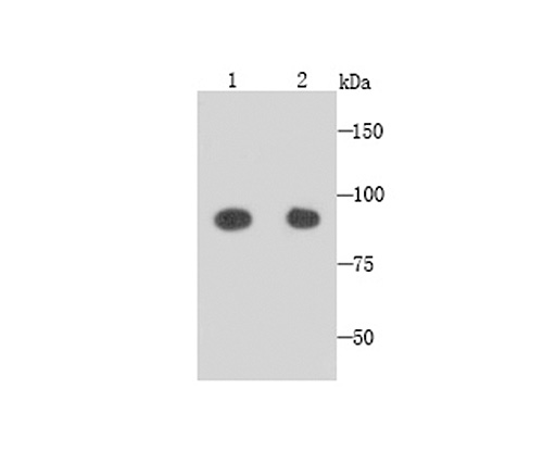
Fig1: Western blot analysis of GRAMD1A on NIH/3T3 (1) and SHG-44 (2) cells lysates using anti-GRAMD1A antibody at 1/1,000 dilution.Application
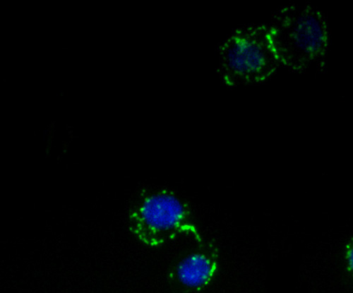
Fig2: ICC staining GRAMD1A in HepG2 cells (green). The nuclear counter stain is DAPI (blue). Cells were fixed in paraformaldehyde, permeabilised with 0.25% Triton X100/PBS.Application
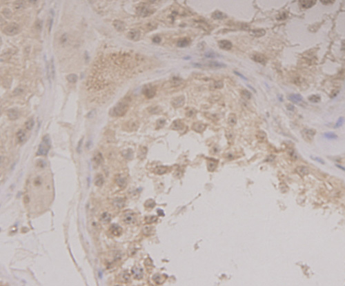
Fig3: Immunohistochemical analysis of paraffin-embedded human tonsil tissue using anti-GRAMD1A antibody. Counter stained with hematoxylin.Application
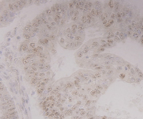
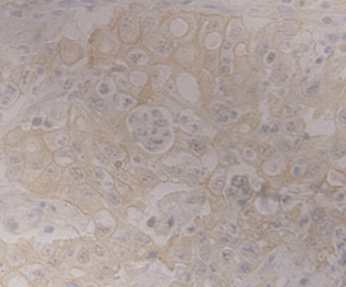
Fig4: Immunohistochemical analysis of paraffin-embedded human endometrial cancer tissue using anti-GRAMD1A antibody. Counter stained with hematoxylin.Application
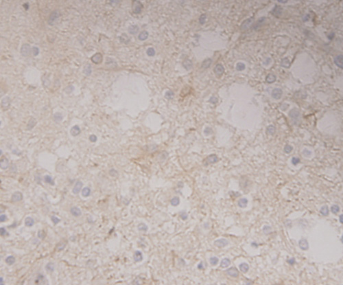
Fig5: Immunohistochemical analysis of paraffin-embedded human glioma tissue using anti-GRAMD1A antibody. Counter stained with hematoxylin.Application
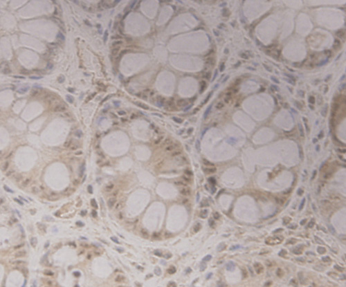
Fig6: Immunohistochemical analysis of paraffin-embedded human colon tissue using anti-GRAMD1A antibody. Counter stained with hematoxylin.Application
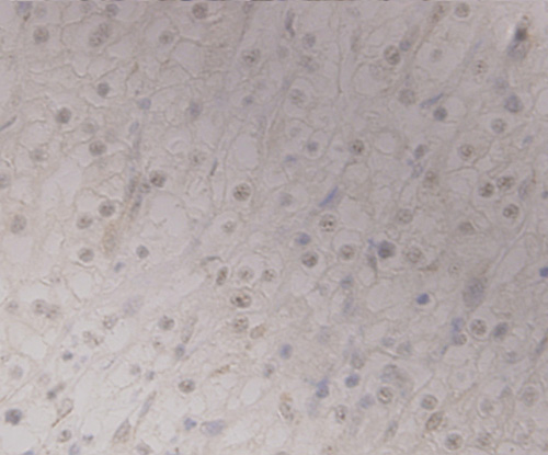
Fig7: Immunohistochemical analysis of paraffin-embedded human renal cell carcinoma tissue using anti-GRAMD1A antibody. Counter stained with hematoxylin.Application
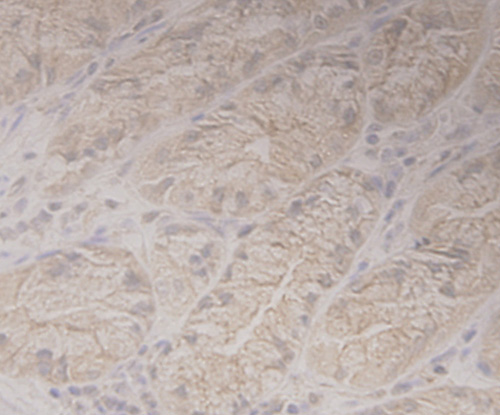
Fig8: Immunohistochemical analysis of paraffin-embedded human stamoch tissue using anti-GRAMD1A antibody. Counter stained with hematoxylin.Application
Fig9: Immunohistochemical analysis of paraffin-embedded human gastric adenocarcinoma tissue using anti-GRAMD1A antibody. Counter stained with hematoxylin.| Product Name | GRAMD1A [1-E4] |
|---|---|
| Antibody Type | Primary Antibodies |
| Product description | GRAMD1A first finds in human embryonic stem cell, and expresses in ectoderm, mesoderm and endoderm tissues and many tumor cells, its function hasn’t been explored. The researh has determined the role of GRAM1DA in the self-renewal of HCC stem cell and resistance to chemotherapy by overexpressing GRAMD1A, and the role of GRAMD1A in tumor growth by overexpressing or downregulating GRAMD1A. GRAMD1A is a prognostic factor for HCC, it promoted the self-renewal of HCC stem cell, resistance to chemotherapy and tumor growth through promoting STAT5, Inhibition of STAT5 in indicated cells with GRAMD1A overexpression inhibited the self-renewal of HCC stem cell, resistance to chemotherapy and tumor growth. |
| Immunogen | Recombinant protein |
| Clonality | Monoclonal |
|---|---|
| Isotype | IgM |
| Host Species | Mouse |
| Tested Applications | WBICCIHC |
| WB:1:250~1:1000 ICC:1:100~1:200 IHC:1:20~1:100 Notes:Optimal dilutions/concentrations should be determined by the researcher. |
|
| Species Reactivity | HumanMouse |
| Concentration | 2mg/ml |
| Molecular Weight(MW) | 80 kDa |
|---|---|
| Cellular Localization | Membrane, nucleus. |
| SwissProt ID | Q96CP6 |
|---|
Application
Fig1: Western blot analysis of GRAMD1A on NIH/3T3 (1) and SHG-44 (2) cells lysates using anti-GRAMD1A antibody at 1/1,000 dilution.
Application
Fig2: ICC staining GRAMD1A in HepG2 cells (green). The nuclear counter stain is DAPI (blue). Cells were fixed in paraformaldehyde, permeabilised with 0.25% Triton X100/PBS.
Application
Fig3: Immunohistochemical analysis of paraffin-embedded human tonsil tissue using anti-GRAMD1A antibody. Counter stained with hematoxylin.
Application
Fig4: Immunohistochemical analysis of paraffin-embedded human endometrial cancer tissue using anti-GRAMD1A antibody. Counter stained with hematoxylin.
Application
Fig5: Immunohistochemical analysis of paraffin-embedded human glioma tissue using anti-GRAMD1A antibody. Counter stained with hematoxylin.
Application
Fig6: Immunohistochemical analysis of paraffin-embedded human colon tissue using anti-GRAMD1A antibody. Counter stained with hematoxylin.
Application
Fig7: Immunohistochemical analysis of paraffin-embedded human renal cell carcinoma tissue using anti-GRAMD1A antibody. Counter stained with hematoxylin.
Application
Fig8: Immunohistochemical analysis of paraffin-embedded human stamoch tissue using anti-GRAMD1A antibody. Counter stained with hematoxylin.
Application
Fig9: Immunohistochemical analysis of paraffin-embedded human gastric adenocarcinoma tissue using anti-GRAMD1A antibody. Counter stained with hematoxylin.| Positive Control | NIH/3T3, SHG-44, HepG2, human tonsil tissue, human endometrial cancer tissue, human glioma tissue, human colon tissue, human renal cell carcinoma tissue, human stamoch tissue, human gastric adenocarcinoma tissue. |
|---|---|
| Application Notes | WB:1:250~1:1000 ICC:1:100~1:200 IHC:1:20~1:100 Notes:Optimal dilutions/concentrations should be determined by the researcher. |
| Form | Liquid |
|---|---|
| Storage Instructions | Store at +4℃ after thawing. Aliquot store at -20℃ or -80℃. Avoid repeated freeze / thaw cycles. |
| Storage Buffer | 1*TBS (pH7.4), 1%BSA, 40%Glycerol. Preservative: 0.05% Sodium Azide |


