Application
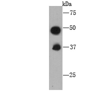
Fig1: Western blot analysis of C10orf58 on recombinant protein using anti-C10orf58 antibody at 1/1000 dilution.Application
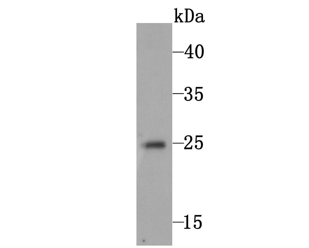
Fig2: Western blot analysis of C10orf58 on human skin tissue lysate using anti-C10orf58 antibody at 1/1000 dilution.Application
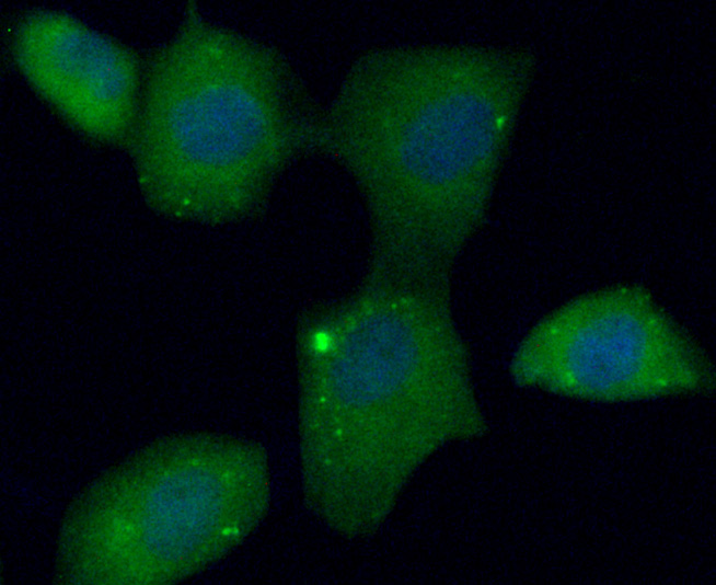
Fig3: ICC staining C10orf58 (green) in HUVEC cells. The nuclear counter stain is DAPI (blue). Cells were fixed in paraformaldehyde, permeabilised with 0.25% Triton X100/PBS.Application
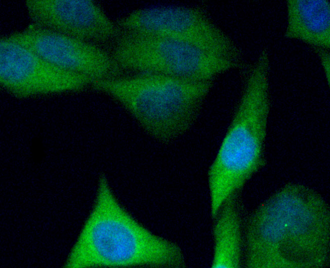
Fig4: ICC staining C10orf58 (green) in PC-3M cells. The nuclear counter stain is DAPI (blue). Cells were fixed in paraformaldehyde, permeabilised with 0.25% Triton X100/PBS.Application
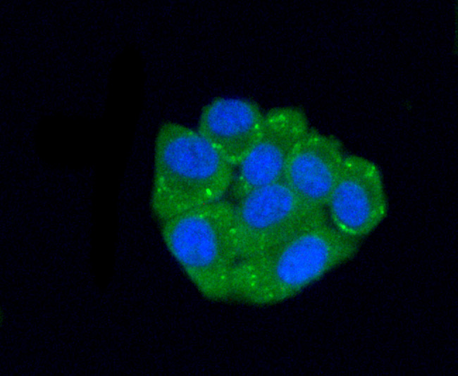
Fig5: ICC staining C10orf58 (green) in HepG2 cells. The nuclear counter stain is DAPI (blue). Cells were fixed in paraformaldehyde, permeabilised with 0.25% Triton X100/PBS.| Product Name | C10orf58 |
|---|---|
| Antibody Type | Primary Antibodies |
| Antigen Alias | C10orf58 antibody CJ058_HUMAN antibody UPF0765 protein C10orf58 antibody |
| Product description | FAM213A, also known as PAMM (peroxiredoxin-like 2 activated in M-CSF stimulated monocytes) or C10orf58, is a 229 amino acid cytoplasmic protein that belongs to the peroxiredoxin-like FAM213 family and FAM213A subfamily. FAM213A plays a role in redox regulation of the cell, acts as an antioxidant, and is induced by CSF1 in peripheral blood mononuclear cells (PBMCs). Existing as two alternatively spliced isoforms, the gene encoding FAM213A maps to human chromosome 10. Spanning nearly 135 million base pairs, chromosome 10 makes up approximately 4.5% of total DNA in cells and encodes nearly 1,200 genes. |
| Immunogen | Recombinant protein |
| Clonality | Monoclonal |
|---|---|
| Isotype | IgG2a |
| Host Species | Mouse |
| Tested Applications | WBICC |
| WB:1:500 ICC:1:50-1:200 |
|
| Species Reactivity | Human |
| Concentration | 2mg/ml |
| Alternative Names | C10orf58 antibody CJ058_HUMAN antibody UPF0765 protein C10orf58 antibody |
|---|---|
| Cellular Localization | Secreted. |
| SwissProt ID | Q9BRX8 |
|---|
Application
Fig1: Western blot analysis of C10orf58 on recombinant protein using anti-C10orf58 antibody at 1/1000 dilution.
Application
Fig2: Western blot analysis of C10orf58 on human skin tissue lysate using anti-C10orf58 antibody at 1/1000 dilution.
Application
Fig3: ICC staining C10orf58 (green) in HUVEC cells. The nuclear counter stain is DAPI (blue). Cells were fixed in paraformaldehyde, permeabilised with 0.25% Triton X100/PBS.
Application
Fig4: ICC staining C10orf58 (green) in PC-3M cells. The nuclear counter stain is DAPI (blue). Cells were fixed in paraformaldehyde, permeabilised with 0.25% Triton X100/PBS.
Application
Fig5: ICC staining C10orf58 (green) in HepG2 cells. The nuclear counter stain is DAPI (blue). Cells were fixed in paraformaldehyde, permeabilised with 0.25% Triton X100/PBS.| Positive Control | Recombinant protein, human skin tissue lysate, HepG2, HUVEC, PC-3M. |
|---|---|
| Application Notes | WB:1:500 ICC:1:50-1:200 |
| Form | Liquid |
|---|---|
| Storage Instructions | Store at +4℃ after thawing. Aliquot store at -20℃ or -80℃. Avoid repeated freeze / thaw cycles. |
| Storage Buffer | 1*TBS (pH7.4), 0.5%BSA, 50%Glycerol. Preservative: 0.05% Sodium Azide. |


