Application
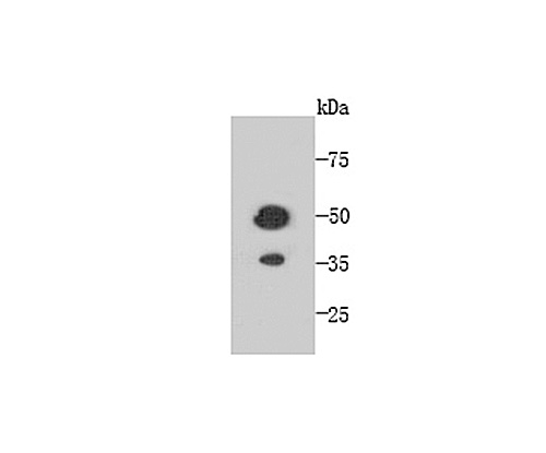
Fig1: Western blot analysis of TMEM177 on recombinant protein using anti-TMEM177 antibody at 1/1,000 dilution.Application
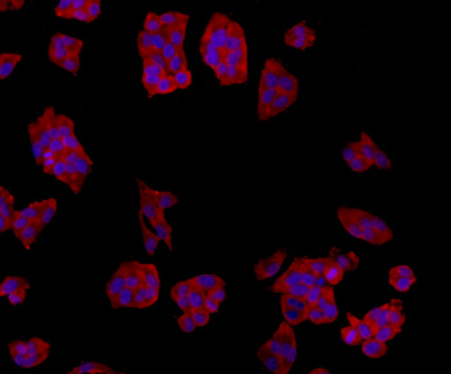
Fig2: ICC staining TMEM177 in Hela cells (red). The nuclear counter stain is DAPI (blue). Cells were fixed in paraformaldehyde, permeabilised with 0.25% Triton X100/PBS.Application
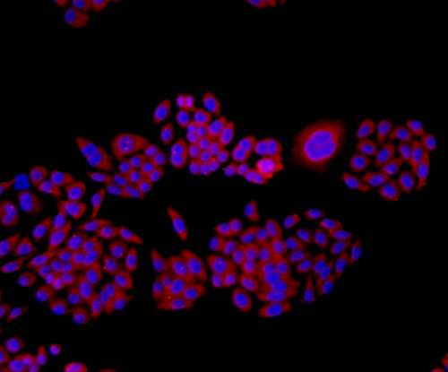
Fig3: ICC staining TMEM177 in HepG2 cells (red). The nuclear counter stain is DAPI (blue). Cells were fixed in paraformaldehyde, permeabilised with 0.25% Triton X100/PBS.Application
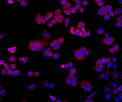
Fig4: ICC staining TMEM177 in MCF-7 cells (red). The nuclear counter stain is DAPI (blue). Cells were fixed in paraformaldehyde, permeabilised with 0.25% Triton X100/PBS.Application
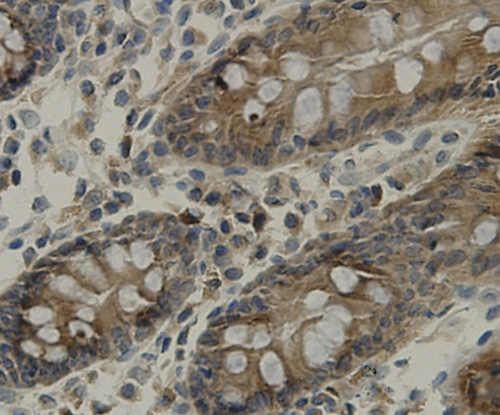
Fig5: Immunohistochemical analysis of paraffin-embedded human colon tissue using anti-TMEM177 antibody. Counter stained with hematoxylin.Application
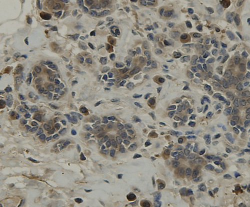
Fig6: Immunohistochemical analysis of paraffin-embedded human breast tissue using anti-TMEM177 antibody. Counter stained with hematoxylin.Application
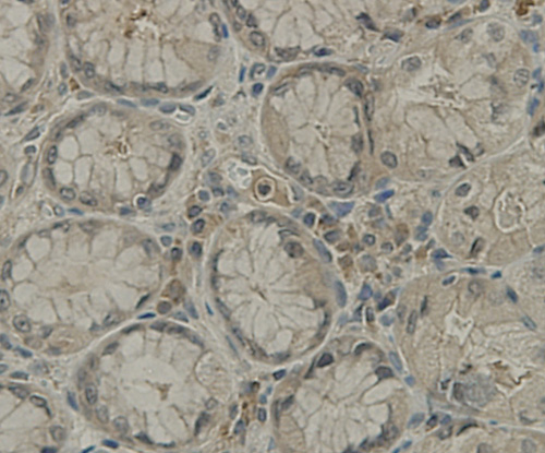
Fig7: Immunohistochemical analysis of paraffin-embedded hman stamoch tissue using anti-TMEM177 antibody. Counter stained with hematoxylin.Application
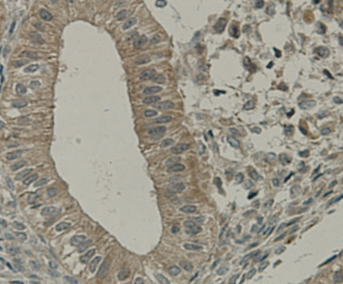
Fig8: Immunohistochemical analysis of paraffin-embedded human uterus muscle tissue using anti-TMEM177 antibody. Counter stained with hematoxylin.| Product Name | TMEM177 [A6-A11-9] |
|---|---|
| Antibody Type | Primary Antibodies |
| Antigen Alias | TM177_HUMAN antibody Tmem177 antibody Transmembrane protein 177 antibody |
| Product description | A transmembrane protein (TMEM) is a type of integral membrane protein that spans the entirety of the biological membrane to which it is permanently attached. Many transmembrane proteins function as gateways to permit the transport of specific substances across the biological membrane. They frequently undergo significant conformational changes to move a substance through the membrane. Transmembrane proteins are polytopic proteins that aggregate and precipitate in water. They require detergents or nonpolar solvents for extraction, although some of them (beta-barrels) can be also extracted using denaturing agents. |
| Immunogen | Recombinant protein |
| Clonality | Monoclonal |
|---|---|
| Isotype | IgG1 |
| Host Species | Mouse |
| Tested Applications | WBICCIHC |
| WB:1:250~1:1000 ICC:1:100~1:200 IHC:1:20~1:100 Notes:Optimal dilutions/concentrations should be determined by the researcher. |
|
| Species Reactivity | Human |
| Concentration | 2mg/ml |
| Alternative Names | TM177_HUMAN antibody Tmem177 antibody Transmembrane protein 177 antibody |
|---|---|
| Molecular Weight(MW) | 34 kDa |
| Cellular Localization | Membrane. |
| SwissProt ID | Q53S58 |
|---|
Application
Fig1: Western blot analysis of TMEM177 on recombinant protein using anti-TMEM177 antibody at 1/1,000 dilution.
Application
Fig2: ICC staining TMEM177 in Hela cells (red). The nuclear counter stain is DAPI (blue). Cells were fixed in paraformaldehyde, permeabilised with 0.25% Triton X100/PBS.
Application
Fig3: ICC staining TMEM177 in HepG2 cells (red). The nuclear counter stain is DAPI (blue). Cells were fixed in paraformaldehyde, permeabilised with 0.25% Triton X100/PBS.
Application
Fig4: ICC staining TMEM177 in MCF-7 cells (red). The nuclear counter stain is DAPI (blue). Cells were fixed in paraformaldehyde, permeabilised with 0.25% Triton X100/PBS.
Application
Fig5: Immunohistochemical analysis of paraffin-embedded human colon tissue using anti-TMEM177 antibody. Counter stained with hematoxylin.
Application
Fig6: Immunohistochemical analysis of paraffin-embedded human breast tissue using anti-TMEM177 antibody. Counter stained with hematoxylin.
Application
Fig7: Immunohistochemical analysis of paraffin-embedded hman stamoch tissue using anti-TMEM177 antibody. Counter stained with hematoxylin.
Application
Fig8: Immunohistochemical analysis of paraffin-embedded human uterus muscle tissue using anti-TMEM177 antibody. Counter stained with hematoxylin.| Positive Control | TMEM177 recombinant protein, HepG2, Hela, MCF-7, human colon tissue, human breast tissue, human stamoch tissue, human uterus tissue. |
|---|---|
| Application Notes | WB:1:250~1:1000 ICC:1:100~1:200 IHC:1:20~1:100 Notes:Optimal dilutions/concentrations should be determined by the researcher. |
| Form | Liquid |
|---|---|
| Storage Instructions | Store at +4℃ after thawing. Aliquot store at -20℃ or -80℃. Avoid repeated freeze / thaw cycles |
| Storage Buffer | 1*TBS (pH7.4), 1%BSA, 40%Glycerol. Preservative: 0.05% Sodium Azide. |


